Vakili Sarah, Ahooyi Taha Mohseni, Yarandi Shadan S, Donadoni Martina, Rappaport Jay, Sariyer Ilker K
Department of Neuroscience and Center for Neurovirology, Temple University Lewis Katz School of Medicine, Philadelphia, PA 19140, USA.
Tulane National Primate Research Center, New Orleans, Covington, LA 70433, USA.
Brain Sci. 2020 Jul 3;10(7):424. doi: 10.3390/brainsci10070424.
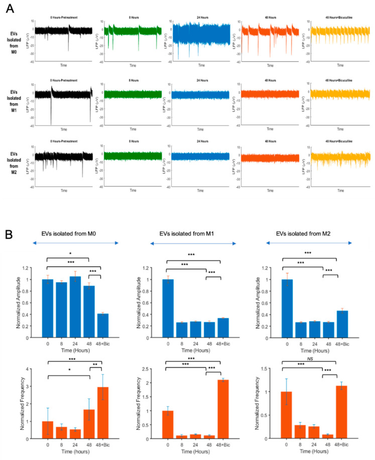
Several factors can contribute to neuroinflammatory disorders, such as cytokine and chemokines that are produced and released from peripherally derived immune cells or from locally activated cells such as microglia and perivascular macrophages in the brain. The primary function of these cells is to clear inflammation; however, following inflammation, circulating monocytes are recruited to the central nervous system (CNS). Monocyte-derived macrophages in the CNS play pivotal roles in mediating neuroinflammatory responses. Macrophages are heterogeneous both in normal and in pathological conditions due to their plasticity, and they are classified in two main subsets, classically activated (M1) or alternatively activated (M2). There is accumulating evidence suggesting that extracellular vesicles (EVs) released from activated immune cells may play crucial roles in mediating inflammation. However, a possible role of EVs released from immune cells such as M1 and M2 macrophages on neuronal functions in the brain is not known. In order to investigate the molecular and cellular impacts of macrophages and EVs released from macrophage subtypes on neuronal functions, we used a recently established in vitro M1 and M2 macrophage culture model and isolated and characterized EVs from these macrophage subtypes, treated primary neurons with M1 or M2 EVs, and analyzed the extracellular action potentials of neurons with microelectrode array studies (MEA). Our results introduce evidence on the interfering role of inflammatory EVs released from macrophages in interneuronal signal transmission processes, with implications in the pathogenesis of neuroinflammatory diseases induced by a variety of inflammatory insults.
多种因素可导致神经炎症性疾病,例如细胞因子和趋化因子,它们由外周来源的免疫细胞产生并释放,或由大脑中局部激活的细胞如小胶质细胞和血管周围巨噬细胞产生并释放。这些细胞的主要功能是清除炎症;然而,炎症发生后,循环单核细胞会被募集到中枢神经系统(CNS)。中枢神经系统中单核细胞衍生的巨噬细胞在介导神经炎症反应中起关键作用。巨噬细胞由于其可塑性,在正常和病理条件下均具有异质性,它们主要分为两个亚群,经典激活的(M1)或交替激活的(M2)。越来越多的证据表明,活化免疫细胞释放的细胞外囊泡(EVs)可能在介导炎症中起关键作用。然而,M1和M2巨噬细胞等免疫细胞释放的EVs对大脑神经元功能的潜在作用尚不清楚。为了研究巨噬细胞及其亚型释放的EVs对神经元功能的分子和细胞影响,我们使用了最近建立的体外M1和M2巨噬细胞培养模型,从这些巨噬细胞亚型中分离并鉴定了EVs,用M1或M2 EVs处理原代神经元,并通过微电极阵列研究(MEA)分析神经元的细胞外动作电位。我们的结果为巨噬细胞释放的炎性EVs在神经元间信号传递过程中的干扰作用提供了证据,这对由各种炎性损伤引起的神经炎症性疾病的发病机制具有重要意义。