Gesperger Johanna, Lichtenegger Antonia, Roetzer Thomas, Salas Matthias, Eugui Pablo, Harper Danielle J, Merkle Conrad W, Augustin Marco, Kiesel Barbara, Mercea Petra A, Widhalm Georg, Baumann Bernhard, Woehrer Adelheid
Center for Medical Physics and Biomedical Engineering, Medical University of Vienna, 1090 Vienna, Austria.
Division of Neuropathology and Neurochemistry, Department of Neurology, Medical University of Vienna, 1090 Vienna, Austria.
Cancers (Basel). 2020 Jul 6;12(7):1806. doi: 10.3390/cancers12071806.
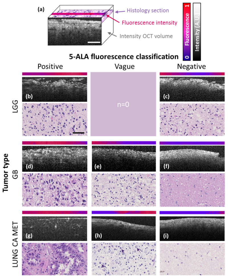
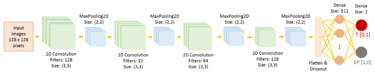
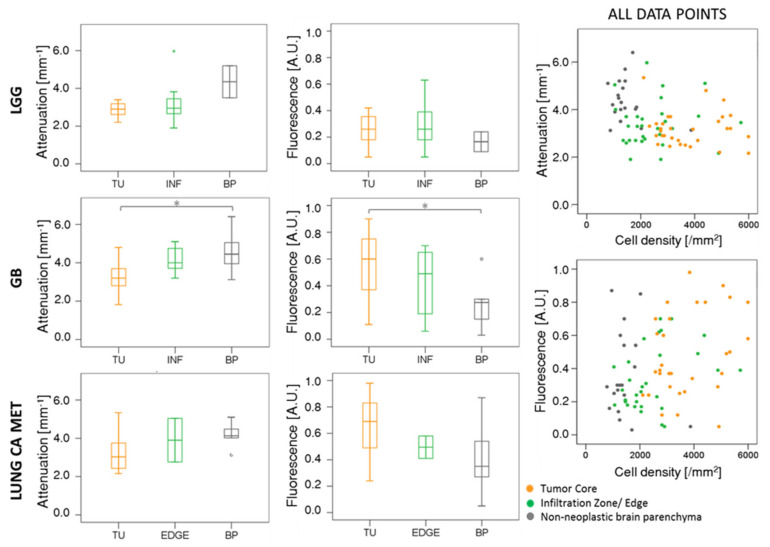
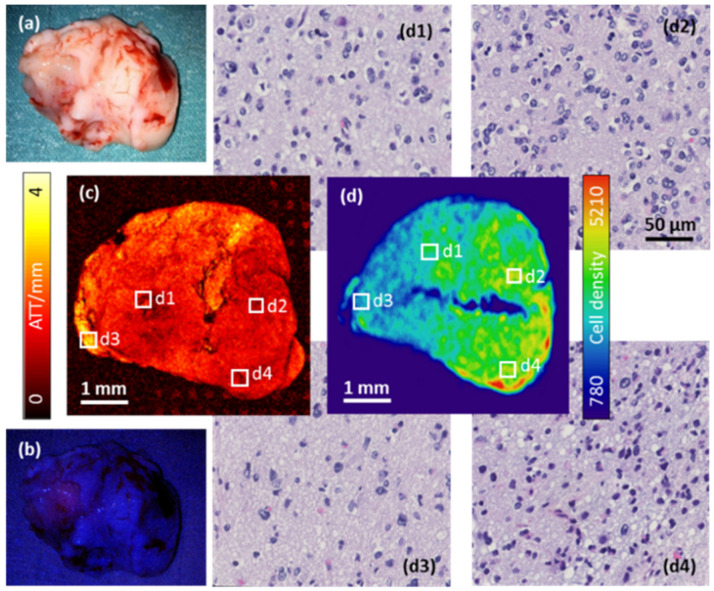
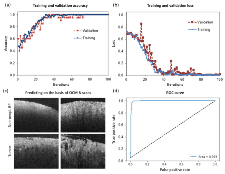
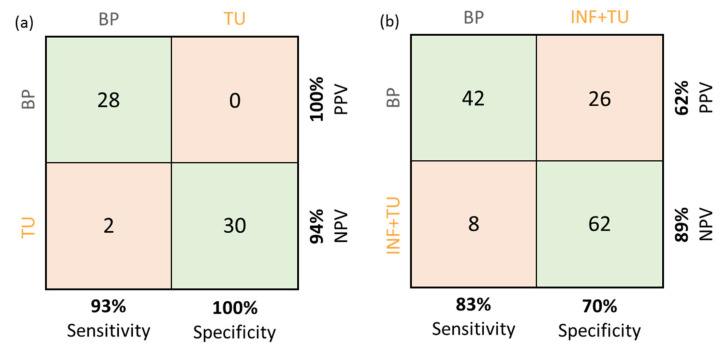
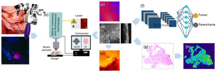
Fluorescence-guided surgery is a state-of-the-art approach for intraoperative imaging during neurosurgical removal of tumor tissue. While the visualization of high-grade gliomas is reliable, lower grade glioma often lack visible fluorescence signals. Here, we present a hybrid prototype combining visible light optical coherence microscopy (OCM) and high-resolution fluorescence imaging for assessment of brain tumor samples acquired by 5-aminolevulinic acid (5-ALA) fluorescence-guided surgery. OCM provides high-resolution information of the inherent tissue scattering and absorption properties of tissue. We here explore quantitative attenuation coefficients derived from volumetric OCM intensity data and quantitative high-resolution 5-ALA fluorescence as potential biomarkers for tissue malignancy including otherwise difficult-to-assess low-grade glioma. We validate our findings against the gold standard histology and use attenuation and fluorescence intensity measures to differentiate between tumor core, infiltrative zone and adjacent brain tissue. Using large field-of-view scans acquired by a near-infrared swept-source optical coherence tomography setup, we provide initial assessments of tumor heterogeneity. Finally, we use cross-sectional OCM images to train a convolutional neural network that discriminates tumor from non-tumor tissue with an accuracy of 97%. Collectively, the present hybrid approach offers potential to translate into an in vivo imaging setup for substantially improved intraoperative guidance of brain tumor surgeries.
荧光引导手术是神经外科手术切除肿瘤组织时进行术中成像的一种先进方法。虽然高级别胶质瘤的可视化是可靠的,但低级别胶质瘤通常缺乏可见的荧光信号。在此,我们展示了一种结合可见光光学相干显微镜(OCM)和高分辨率荧光成像的混合原型,用于评估通过5-氨基乙酰丙酸(5-ALA)荧光引导手术获取的脑肿瘤样本。OCM提供了组织固有散射和吸收特性的高分辨率信息。我们在此探索从体积OCM强度数据得出的定量衰减系数以及定量高分辨率5-ALA荧光,作为包括难以评估的低级别胶质瘤在内的组织恶性程度的潜在生物标志物。我们对照金标准组织学验证了我们的发现,并使用衰减和荧光强度测量来区分肿瘤核心、浸润区和相邻脑组织。通过使用近红外扫频源光学相干断层扫描设备获取的大视野扫描,我们对肿瘤异质性进行了初步评估。最后,我们使用横截面OCM图像训练了一个卷积神经网络,该网络区分肿瘤和非肿瘤组织的准确率为97%。总体而言,目前的混合方法有可能转化为一种体内成像设置,以显著改善脑肿瘤手术的术中引导。