Department of Neurosurgery, University of Michigan, Ann Arbor, MI, USA.
School of Medicine, University of Michigan, Ann Arbor, MI, USA.
Nat Med. 2020 Jan;26(1):52-58. doi: 10.1038/s41591-019-0715-9. Epub 2020 Jan 6.
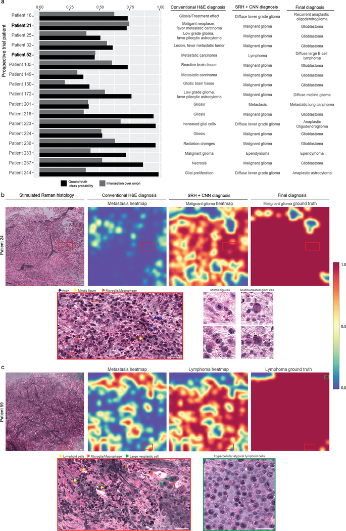
Intraoperative diagnosis is essential for providing safe and effective care during cancer surgery. The existing workflow for intraoperative diagnosis based on hematoxylin and eosin staining of processed tissue is time, resource and labor intensive. Moreover, interpretation of intraoperative histologic images is dependent on a contracting, unevenly distributed, pathology workforce. In the present study, we report a parallel workflow that combines stimulated Raman histology (SRH), a label-free optical imaging method and deep convolutional neural networks (CNNs) to predict diagnosis at the bedside in near real-time in an automated fashion. Specifically, our CNNs, trained on over 2.5 million SRH images, predict brain tumor diagnosis in the operating room in under 150 s, an order of magnitude faster than conventional techniques (for example, 20-30 min). In a multicenter, prospective clinical trial (n = 278), we demonstrated that CNN-based diagnosis of SRH images was noninferior to pathologist-based interpretation of conventional histologic images (overall accuracy, 94.6% versus 93.9%). Our CNNs learned a hierarchy of recognizable histologic feature representations to classify the major histopathologic classes of brain tumors. In addition, we implemented a semantic segmentation method to identify tumor-infiltrated diagnostic regions within SRH images. These results demonstrate how intraoperative cancer diagnosis can be streamlined, creating a complementary pathway for tissue diagnosis that is independent of a traditional pathology laboratory.
术中诊断对于癌症手术期间提供安全有效的护理至关重要。基于处理组织的苏木精和伊红染色的现有术中诊断工作流程既费时、资源密集又耗费劳动力。此外,术中组织学图像的解释依赖于数量有限、分布不均的病理科劳动力。在本研究中,我们报告了一种平行工作流程,该流程结合了无标记的光学成像方法刺激拉曼组织学(SRH)和深度卷积神经网络(CNN),以自动化方式在接近实时的情况下预测床边诊断。具体来说,我们的 CNN 经过超过 250 万张 SRH 图像的训练,可以在 150 秒内预测手术室中的脑瘤诊断,比传统技术快一个数量级(例如,20-30 分钟)。在一项多中心前瞻性临床试验(n=278)中,我们证明了基于 CNN 的 SRH 图像诊断与病理学家基于传统组织学图像的解释具有非劣效性(总体准确性为 94.6%对 93.9%)。我们的 CNN 学习了可识别的组织学特征表示层次结构,以对脑肿瘤的主要组织病理学类型进行分类。此外,我们还实施了一种语义分割方法来识别 SRH 图像中的肿瘤浸润诊断区域。这些结果表明,术中癌症诊断如何实现流程化,为组织诊断创建了一种独立于传统病理实验室的互补途径。