Mansour Amr, Elfiky Azza Abdallah, Mohamed Alaa Sakran, Ezzeldin Dina Adel
Department of Cardiology, Ain Shams University Hospital, Cairo, Egypt.
Department of Cardiology, Dar Al Fouad Hospital, Cairo, Egypt.
Ann Pediatr Cardiol. 2020 Apr-Jun;13(2):123-129. doi: 10.4103/apc.APC_93_18. Epub 2020 Mar 20.
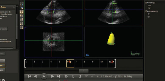
The main effect of pulmonary stenosis is a rise in right ventricular pressure. This pressure overload leads to multiple changes in the shape, dimensions, and volumes of the right ventricle (RV) that are reversed after the relieve of the valve obstruction. We thought to study the changes in the RV in patients undergoing balloon pulmonary valvuloplasty (BPV) using three-dimensional (3D) echocardiography.
The study included 50 patients with isolated valvular pulmonary stenosis who underwent BPV at our hospital from December 2016 to August 2017; echocardiography was recorded preprocedural and 3 months after the procedural.
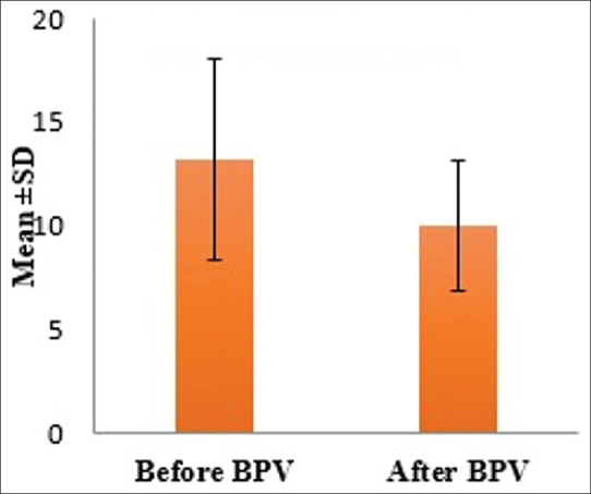
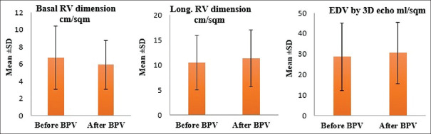
The median age of the study group at the time of the procedure was 2.7 years. The indexed RV wall thickness, basal, and mid-right ventricular dimensions decreased significantly after the procedure ( < 0.005), and the longitudinal dimension increased significantly after the procedure ( < 0.005). The end-systolic and the end-diastolic volumes (EDVs) by 3D echocardiography increased insignificantly ( > 0.05), and the right ventricular function increased significantly ( < 0.05), indicating that the changes in the EDVs were more than the changes in the end-systolic volumes.
There are several factors that interplay together and result in reverse remodeling of the RV after BPV including regression in the RV hypertrophy; changes in the interventricular septal morphology, bowing, and mobility; and changes in the ventricular geometry and dimensions, rather than changes in the ventricular volumes.
肺动脉狭窄的主要影响是右心室压力升高。这种压力超负荷会导致右心室(RV)的形状、尺寸和容积发生多种变化,而在瓣膜梗阻解除后这些变化会逆转。我们想用三维(3D)超声心动图研究接受球囊肺动脉瓣成形术(BPV)患者的右心室变化。
该研究纳入了2016年12月至2017年8月在我院接受BPV的50例单纯瓣膜性肺动脉狭窄患者;在术前和术后3个月记录超声心动图。
研究组手术时的中位年龄为2.7岁。术后右心室壁厚度指数、基底及右心室中部尺寸显著减小(<0.005),纵向尺寸术后显著增加(<0.005)。三维超声心动图测得的收缩末期和舒张末期容积(EDV)增加不显著(>0.05),右心室功能显著增加(<0.05),表明舒张末期容积的变化大于收缩末期容积的变化。
有几个因素相互作用,导致BPV后右心室发生逆向重构,包括右心室肥厚的消退;室间隔形态、弯曲度和活动度的变化;以及心室几何形状和尺寸的变化,而非心室容积的变化。