Wellcome Centre for Mitochondrial Research, Newcastle University, Newcastle upon Tyne, UK.
Translational and Clinical Research Institute, Newcastle University, Newcastle upon Tyne, UK.
Acta Neuropathol Commun. 2020 Jul 9;8(1):103. doi: 10.1186/s40478-020-00985-8.
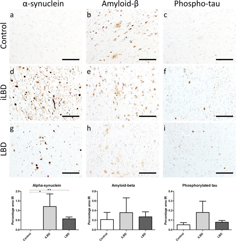
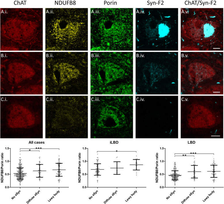
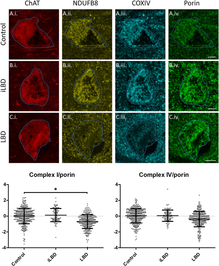
Neurons of the nucleus basalis of Meynert (nbM) are vulnerable to Lewy body formation and neuronal loss, which is thought to underlie cognitive dysfunction in Lewy body dementia (LBD). There is continued debate about whether Lewy bodies exert a neurodegenerative effect by affecting mitochondria, or whether they represent a protective mechanism. Therefore, the present study sought to determine whether the nbM is subject to mitochondrial dysfunctional in LBD and the association of Lewy body formation with such changes. Post-mortem nbM tissue was stained for Complex I or IV and quantitated relative to porin with immunofluorescence using confocal microscopy of individual cells from LBD (303 neurons, 8 cases), control (362 neurons, 8 cases) and asymptomatic incidental LBD (iLBD) cases (99 neurons, 2 cases). Additionally, α-synuclein, tau and amyloid-β pathology were analysed using quantitative immunohistochemistry, and respiratory chain markers were compared in cells with Lewy bodies (N = 134) and unaffected cells (N = 272). The expression of Complex I normalised to mitochondrial mass was significantly lower in LBD compared to control and iLBD cases and this was unrelated to local neuropathological burdens but trended toward a relationship with neuronal loss. Furthermore, Complex I expression was higher in cells with Lewy bodies compared to adjacent cells without α-synuclein aggregates. These findings suggest that Complex I deficits in the nbM occur in symptomatic LBD cases and may relate to neuronal loss, but that contrary to the view that Lewy body formation underlies neuronal dysfunction and damage in LBD, Lewy bodies are associated with higher Complex I expression than neurons without Lewy bodies. One could speculate that Lewy bodies may provide a mechanism to encapsulate damaged mitochondria and/or α-synuclein oligomers, thus protecting neurons from their cytotoxic effects.
基底核内梅内尔特核(nbM)的神经元易受路易体形成和神经元丢失的影响,这被认为是路易体痴呆(LBD)认知功能障碍的基础。目前仍在争论路易体是否通过影响线粒体发挥神经退行性作用,还是它们代表一种保护机制。因此,本研究旨在确定 LBD 中 nbM 是否存在线粒体功能障碍,以及路易体形成与这种变化的关系。使用共聚焦显微镜对来自 LBD(303 个神经元,8 例)、对照(362 个神经元,8 例)和无症状偶发性 LBD(iLBD)病例(99 个神经元,2 例)的单个细胞进行免疫荧光染色,检测 nbM 组织中复合物 I 或 IV 的染色,并相对于孔蛋白进行定量。此外,使用定量免疫组织化学分析α-突触核蛋白、tau 和淀粉样β病理学,并比较具有路易体(N=134)和无路易体(N=272)的细胞中的呼吸链标志物。与对照和 iLBD 病例相比,LBD 中复合物 I 的表达归一化到线粒体质量显著降低,这与局部神经病理学负担无关,但与神经元丢失呈趋势相关。此外,与相邻无α-突触核蛋白聚集的细胞相比,具有路易体的细胞中复合物 I 的表达更高。这些发现表明,nbM 中的复合物 I 缺陷发生在有症状的 LBD 病例中,可能与神经元丢失有关,但与路易体形成是 LBD 中神经元功能障碍和损伤的基础这一观点相反,路易体与无路易体的神经元相比,表达更高的复合物 I。人们可以推测,路易体可能提供了一种机制来包裹受损的线粒体和/或α-突触核蛋白寡聚物,从而保护神经元免受其细胞毒性作用。