• 文献检索
  • 文档翻译
  • 深度研究
  • 学术资讯
  • Suppr Zotero 插件Zotero 插件
  • 邀请有礼
  • 套餐&价格
  • 历史记录
应用&插件
Suppr Zotero 插件Zotero 插件浏览器插件Mac 客户端Windows 客户端微信小程序
定价
高级版会员购买积分包购买API积分包
服务
文献检索文档翻译深度研究API 文档MCP 服务
关于我们
关于 Suppr公司介绍联系我们用户协议隐私条款
关注我们

Suppr 超能文献

核心技术专利:CN118964589B侵权必究
粤ICP备2023148730 号-1Suppr @ 2026

文献检索

告别复杂PubMed语法,用中文像聊天一样搜索,搜遍4000万医学文献。AI智能推荐,让科研检索更轻松。

立即免费搜索

文件翻译

保留排版,准确专业,支持PDF/Word/PPT等文件格式,支持 12+语言互译。

免费翻译文档

深度研究

AI帮你快速写综述,25分钟生成高质量综述,智能提取关键信息,辅助科研写作。

立即免费体验

利用耐锇远红蛋白通过树脂内 CLEM 对细胞质细胞器进行可视化。

Visualization of cytoplasmic organelles via in-resin CLEM using an osmium-resistant far-red protein.

机构信息

Department of Cellular and Molecular Neuropathology, Juntendo University Graduate School of Medicine, Tokyo, Japan.

Laboratory of Morphology and Image Analysis, Research Support Center, Juntendo University Graduate School of Medicine, Tokyo, Japan.

出版信息

Sci Rep. 2020 Jul 9;10(1):11314. doi: 10.1038/s41598-020-68191-z.

DOI:10.1038/s41598-020-68191-z
PMID:32647231
原文链接:https://pmc.ncbi.nlm.nih.gov/articles/PMC7347593/
Abstract

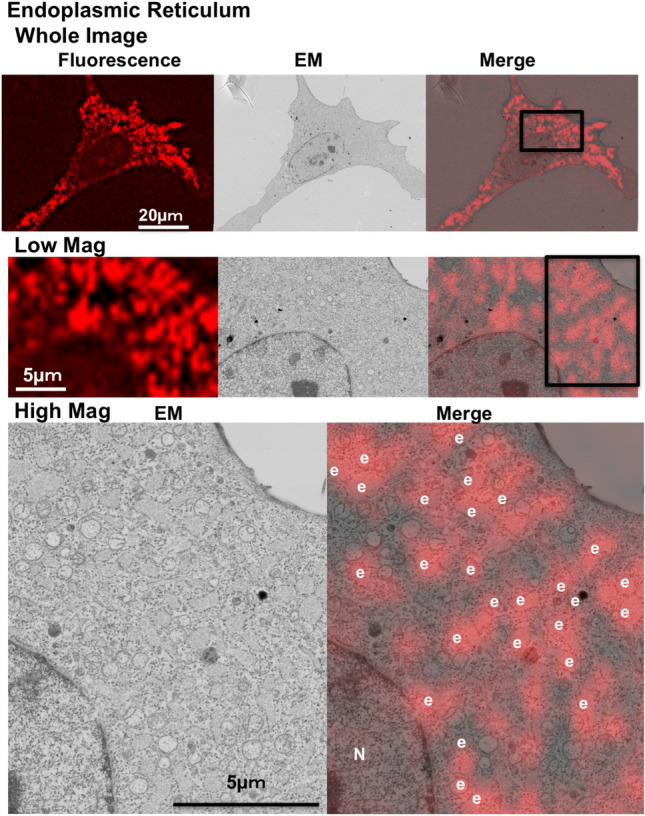
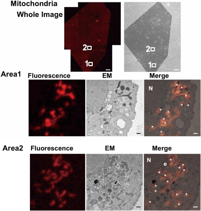
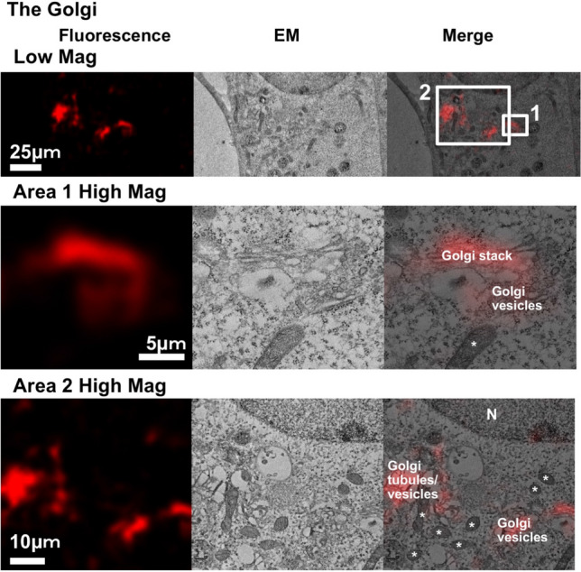
Post-fixation with osmium tetroxide staining and the embedding of Epon are robust and essential treatments that are used to preserve and visualize intracellular membranous structures during electron microscopic analyses. These treatments, however, can significantly diminish the fluorescent intensity of most fluorescent proteins in cells, which creates an obstacle for the in-resin correlative light-electron microscopy (CLEM) of Epon-embedded cells. In this study, we used a far-red fluorescent protein that retains fluorescence after osmium staining and Epon embedding to perform an in-resin CLEM of Epon-embedded samples. The fluorescence of this protein was detected in 100 nm thin sections of the cells in Epon-embedded samples after fixation with 2.5% glutaraldehyde and post-fixation with 1% osmium tetroxide. We performed in-resin CLEM of the mitochondria in Epon-embedded cells using a mitochondria-localized fluorescent protein. Using this protein, we achieved in-resin CLEM of the Golgi apparatus and the endoplasmic reticulum in thin sections of the cells in Epon-embedded samples. To our knowledge, this is the first reported use of a far-red fluorescent protein retains its fluorescence after osmium staining and Epon-embedding, and it represents the first achievement of in-resin CLEM of both the Golgi apparatus and the endoplasmic reticulum in Epon-embedded samples.

摘要

锇酸后固定和环氧树脂包埋是强有力的和必不可少的处理方法,用于在电子显微镜分析中保存和可视化细胞内的膜结构。然而,这些处理方法会显著降低细胞中大多数荧光蛋白的荧光强度,这为环氧树脂包埋细胞的在树脂中相关的光电子显微镜(CLEM)分析带来了障碍。在本研究中,我们使用了一种远红色荧光蛋白,它在锇染色和环氧树脂包埋后仍保留荧光,从而对环氧树脂包埋的样品进行在树脂中 CLEM。该蛋白的荧光在经 2.5%戊二醛固定和 1%锇酸后固定的环氧树脂包埋样品的细胞 100nm 薄片中被检测到。我们使用定位于线粒体的荧光蛋白对环氧树脂包埋细胞中的线粒体进行了在树脂中 CLEM。使用这种蛋白,我们在环氧树脂包埋样品的细胞薄片中实现了高尔基体和内质网的在树脂中 CLEM。据我们所知,这是第一个报道的远红色荧光蛋白在锇染色和环氧树脂包埋后仍保留其荧光的例子,也是第一个在环氧树脂包埋样品中实现高尔基体和内质网的在树脂中 CLEM 的例子。

https://cdn.ncbi.nlm.nih.gov/pmc/blobs/7cd0/7347593/48397f1aea61/41598_2020_68191_Fig4_HTML.jpg
https://cdn.ncbi.nlm.nih.gov/pmc/blobs/7cd0/7347593/00dcaff84fb3/41598_2020_68191_Fig1_HTML.jpg
https://cdn.ncbi.nlm.nih.gov/pmc/blobs/7cd0/7347593/d5c34187fab9/41598_2020_68191_Fig2_HTML.jpg
https://cdn.ncbi.nlm.nih.gov/pmc/blobs/7cd0/7347593/9d967f2491e2/41598_2020_68191_Fig3_HTML.jpg
https://cdn.ncbi.nlm.nih.gov/pmc/blobs/7cd0/7347593/48397f1aea61/41598_2020_68191_Fig4_HTML.jpg

相似文献

1
Visualization of cytoplasmic organelles via in-resin CLEM using an osmium-resistant far-red protein.利用耐锇远红蛋白通过树脂内 CLEM 对细胞质细胞器进行可视化。
Sci Rep. 2020 Jul 9;10(1):11314. doi: 10.1038/s41598-020-68191-z.
2
Osmium-Resistant Fluorescent Proteins and In-Resin Correlative Light-Electron Microscopy of Epon-Embedded Mammalian Cultured Cells.锇抗荧光蛋白和哺乳动物培养细胞包埋后树脂的共相关光电子显微镜。
Methods Mol Biol. 2023;2564:287-297. doi: 10.1007/978-1-0716-2667-2_15.
3
Two-color in-resin CLEM of Epon-embedded cells using osmium resistant green and red fluorescent proteins.利用耐锇的绿色和红色荧光蛋白对包埋在环氧树脂中的细胞进行双色在树脂内 CLEM。
Sci Rep. 2020 Dec 14;10(1):21871. doi: 10.1038/s41598-020-78879-x.
4
Application of immuno- and affinity labeling with fluorescent dyes to in-resin CLEM of Epon-embedded cells.免疫和荧光染料亲和标记在环氧树脂包埋细胞的树脂内免疫电镜中的应用。
Heliyon. 2023 Jun 17;9(6):e17394. doi: 10.1016/j.heliyon.2023.e17394. eCollection 2023 Jun.
5
In-resin CLEM of Epon-embedded cells using proximity labeling.利用邻近标记进行包埋细胞的树脂内 CLEM。
Sci Rep. 2022 Jul 1;12(1):11130. doi: 10.1038/s41598-022-15438-6.
6
Recent advances in in-resin correlative light and electron microscopy of Epon-embedded cells.包埋细胞内明场和电子显微镜关联技术的最新进展
Microscopy (Oxf). 2023 Oct 9;72(5):383-387. doi: 10.1093/jmicro/dfad028.
7
mEosEM withstands osmium staining and Epon embedding for super-resolution CLEM.mEosEM 可耐受锇染色和环氧树脂包埋用于超分辨率 CLEM。
Nat Methods. 2020 Jan;17(1):55-58. doi: 10.1038/s41592-019-0613-6. Epub 2019 Oct 14.
8
Rapid in-EPON CLEM: Combining fast and efficient labeling of self-labeling enzyme tags with EM-resistant Janelia Fluor dyes and StayGold.快速原位光镜-电镜关联技术:将自标记酶标签与抗电子显微镜的詹妮莉荧光染料及StayGold进行快速高效标记相结合。
Heliyon. 2024 Mar 18;10(7):e28055. doi: 10.1016/j.heliyon.2024.e28055. eCollection 2024 Apr 15.
9
A simple preparation method for CLEM using pre-embedding immunohistochemistry with a novel fluorescent probe and stable embedding resin.一种使用新型荧光探针和稳定包埋树脂进行预包埋免疫组织化学的CLEM简单制备方法。
Microscopy (Oxf). 2021 Aug 9;70(4):368-374. doi: 10.1093/jmicro/dfab005.
10
Three-dimensional analysis of the intracellular architecture by scanning electron microscopy.通过扫描电子显微镜对细胞内结构进行三维分析。
Microscopy (Oxf). 2024 Jun 6;73(3):215-225. doi: 10.1093/jmicro/dfad050.

引用本文的文献

1
Introduction to the CLEM technique developed in the field of neuroanatomy.神经解剖学领域中开发的CLEM技术介绍。
Anat Sci Int. 2025 Jul 23. doi: 10.1007/s12565-025-00875-w.
2
α-Synuclein-Assembled Synaptic Vesicle Pools at the Presynaptic Terminal: A Study of α-Synuclein Function Using a Novel Mouse Model.突触前末端的α-突触核蛋白组装的突触小泡池:使用新型小鼠模型对α-突触核蛋白功能的研究
Acta Histochem Cytochem. 2025 Jun 24;58(3):107-114. doi: 10.1267/ahc.25-00017. Epub 2025 Jun 18.
3
A highly stable monomeric red fluorescent protein for advanced microscopy.

本文引用的文献

1
mEosEM withstands osmium staining and Epon embedding for super-resolution CLEM.mEosEM 可耐受锇染色和环氧树脂包埋用于超分辨率 CLEM。
Nat Methods. 2020 Jan;17(1):55-58. doi: 10.1038/s41592-019-0613-6. Epub 2019 Oct 14.
2
Optimization of mNeonGreen for Homo sapiens increases its fluorescent intensity in mammalian cells.针对智人的mNeonGreen优化提高了其在哺乳动物细胞中的荧光强度。
PLoS One. 2018 Jan 17;13(1):e0191108. doi: 10.1371/journal.pone.0191108. eCollection 2018.
3
Correlative In-Resin Super-Resolution Fluorescence and Electron Microscopy of Cultured Cells.
一种用于先进显微镜技术的高度稳定的单体红色荧光蛋白。
Nat Methods. 2025 Apr 17. doi: 10.1038/s41592-025-02676-5.
4
Far-Red Fluorescent Proteins: Tools for Advancing In Vivo Imaging.远红荧光蛋白:活体成像的工具。
Biosensors (Basel). 2024 Jul 24;14(8):359. doi: 10.3390/bios14080359.
5
Rapid in-EPON CLEM: Combining fast and efficient labeling of self-labeling enzyme tags with EM-resistant Janelia Fluor dyes and StayGold.快速原位光镜-电镜关联技术:将自标记酶标签与抗电子显微镜的詹妮莉荧光染料及StayGold进行快速高效标记相结合。
Heliyon. 2024 Mar 18;10(7):e28055. doi: 10.1016/j.heliyon.2024.e28055. eCollection 2024 Apr 15.
6
Application of immuno- and affinity labeling with fluorescent dyes to in-resin CLEM of Epon-embedded cells.免疫和荧光染料亲和标记在环氧树脂包埋细胞的树脂内免疫电镜中的应用。
Heliyon. 2023 Jun 17;9(6):e17394. doi: 10.1016/j.heliyon.2023.e17394. eCollection 2023 Jun.
7
Recent technological advances in correlative light and electron microscopy for the comprehensive analysis of neural circuits.用于神经回路综合分析的相关光电子显微镜技术的最新进展。
Front Neuroanat. 2022 Nov 30;16:1061078. doi: 10.3389/fnana.2022.1061078. eCollection 2022.
8
In-resin CLEM of Epon-embedded cells using proximity labeling.利用邻近标记进行包埋细胞的树脂内 CLEM。
Sci Rep. 2022 Jul 1;12(1):11130. doi: 10.1038/s41598-022-15438-6.
9
Correlative Organelle Microscopy: Fluorescence Guided Volume Electron Microscopy of Intracellular Processes.相关细胞器显微镜检查:细胞内过程的荧光引导体积电子显微镜检查
Front Cell Dev Biol. 2022 Apr 11;10:829545. doi: 10.3389/fcell.2022.829545. eCollection 2022.
10
Improved Fluorescent Proteins for Dual-Colour Post-Embedding CLEM.用于双标记后嵌入 CLEM 的改良荧光蛋白。
Cells. 2022 Mar 23;11(7):1077. doi: 10.3390/cells11071077.
培养细胞的相关树脂内超分辨率荧光和电子显微镜技术
Methods Mol Biol. 2017;1663:163-177. doi: 10.1007/978-1-4939-7265-4_14.
4
Correlative super-resolution fluorescence and electron microscopy using conventional fluorescent proteins in vacuo.在真空中使用传统荧光蛋白的相关超分辨率荧光和电子显微镜技术。
J Struct Biol. 2017 Aug;199(2):120-131. doi: 10.1016/j.jsb.2017.05.013. Epub 2017 May 30.
5
Diverse protocols for correlative super-resolution fluorescence imaging and electron microscopy of chemically fixed samples.用于化学固定样本的相关超分辨率荧光成像和电子显微镜的多种方案。
Nat Protoc. 2017 May;12(5):916-946. doi: 10.1038/nprot.2017.017. Epub 2017 Apr 6.
6
Cysteine Sulfoxidation Increases the Photostability of Red Fluorescent Proteins.半胱氨酸硫氧化增强红色荧光蛋白的光稳定性。
ACS Chem Biol. 2016 Oct 21;11(10):2679-2684. doi: 10.1021/acschembio.6b00579. Epub 2016 Sep 12.
7
Correlative in-resin super-resolution and electron microscopy using standard fluorescent proteins.使用标准荧光蛋白的相关树脂内超分辨率和电子显微镜技术。
Sci Rep. 2015 Mar 31;5:9583. doi: 10.1038/srep09583.
8
Fixation-resistant photoactivatable fluorescent proteins for CLEM.用于CLEM的抗固定光激活荧光蛋白。
Nat Methods. 2015 Mar;12(3):215-8, 4 p following 218. doi: 10.1038/nmeth.3225. Epub 2015 Jan 12.
9
Engineering an improved light-induced dimer (iLID) for controlling the localization and activity of signaling proteins.设计一种改进的光诱导二聚体(iLID)以控制信号蛋白的定位和活性。
Proc Natl Acad Sci U S A. 2015 Jan 6;112(1):112-7. doi: 10.1073/pnas.1417910112. Epub 2014 Dec 22.
10
Correlative light and electron microscopy of GFP.绿色荧光蛋白的相关光镜和电镜检查
Methods Cell Biol. 2012;111:117-38. doi: 10.1016/B978-0-12-416026-2.00007-8.