Song Kyu Young, Desar Sabina, Pengo Thomas, Shanley Ryan, Giubellino Alessio
Department of Laboratory Medicine and Pathology, University of Minnesota, Minneapolis, MN 55455, USA.
Masonic Cancer Center, University of Minnesota, Minneapolis, MN 55455, USA.
Cancers (Basel). 2020 Jul 9;12(7):1847. doi: 10.3390/cancers12071847.
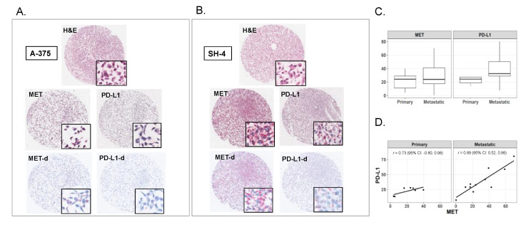
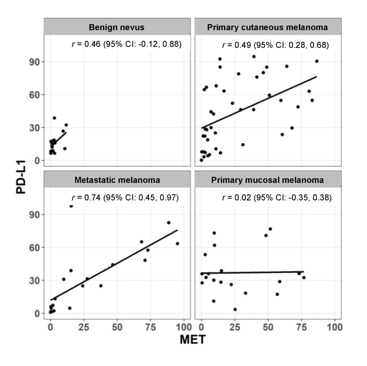
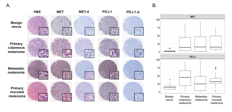
The proto-oncogene MET, the hepatocyte growth factor (HGF) receptor, is a transmembrane receptor tyrosine kinase (RTK) with a prominent role in tumor metastasis and resistance to anti-cancer therapies. Melanoma demonstrates relatively frequent MET aberrations, including MET gene amplification. Concurrently, programmed death-ligand 1 (PD-L1), with its ability to evade anti-tumor immune responses, has emerged as a prominent therapeutic target in melanoma and other malignancies and its expression is used as a predictive biomarker of response to immunotherapy. We performed immunohistochemistry analysis of MET and PD-L1 in 18 human melanoma cell lines derived from both primary and metastatic lesions, and in a human melanoma tissue microarray containing one hundreds melanocytic lesions, including primary cutaneous melanomas, primary mucosal melanomas, metastatic melanomas and benign melanocytic nevi as controls. After color deconvolution, each core was segmented to isolate staining and calculate the percentage of positive cells. Overall, MET expression was higher in tumors with increased PD-L1 expression. Moreover, a robust correlation between MET and PD-L1 expression was found in samples from metastatic melanoma and not in primary cutaneous or mucosal melanoma. These data suggest that relative expression levels of these proteins in combination is a marker of advanced disease and testing for expression of these markers should be considered in patients with melanoma.
原癌基因MET是肝细胞生长因子(HGF)受体,是一种跨膜受体酪氨酸激酶(RTK),在肿瘤转移和抗癌治疗耐药性方面发挥着重要作用。黑色素瘤中MET异常较为常见,包括MET基因扩增。同时,程序性死亡配体1(PD-L1)具有逃避抗肿瘤免疫反应的能力,已成为黑色素瘤和其他恶性肿瘤的重要治疗靶点,其表达被用作免疫治疗反应的预测生物标志物。我们对来自原发性和转移性病变的18个人类黑色素瘤细胞系以及包含100个黑素细胞病变的人类黑色素瘤组织芯片进行了MET和PD-L1的免疫组织化学分析,其中包括原发性皮肤黑色素瘤、原发性黏膜黑色素瘤、转移性黑色素瘤和作为对照的良性黑素细胞痣。经过颜色反卷积后,对每个核心进行分割以分离染色并计算阳性细胞百分比。总体而言,在PD-L1表达增加的肿瘤中MET表达更高。此外,在转移性黑色素瘤样本中发现MET和PD-L1表达之间存在强相关性,而在原发性皮肤或黏膜黑色素瘤中则未发现。这些数据表明,这些蛋白的相对表达水平联合起来是晚期疾病的标志物,对于黑色素瘤患者应考虑检测这些标志物的表达。