Edward S. Harkness Eye Institute, Department of Ophthalmology, Columbia University Irving Medical Center, New York-Presbyterian Hospital, 635 W 165th St, New York, NY, 10032, USA.
Department of Ophthalmology, New York University School of Medicine, New York University Langone Health, New York, NY, USA.
BMC Ophthalmol. 2020 Jul 13;20(1):283. doi: 10.1186/s12886-020-01540-8.
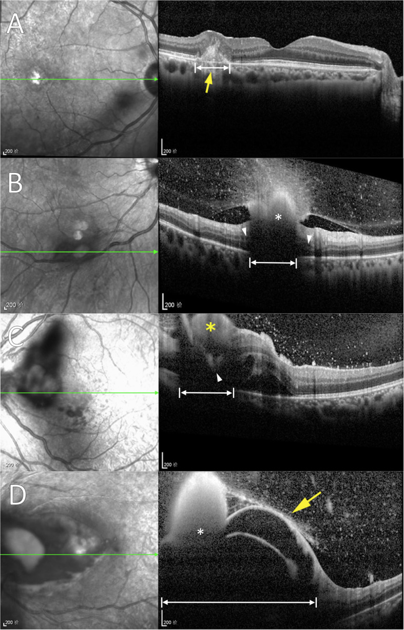
Endogenous Candida endophthalmitis (ECE) has been established with microscopic histopathology, both by autopsy and experimentation, to primarily originate from and involve the choroid. Zhuang et al. examined a series of patients with ECE using spectral-domain optical coherence tomography (SD-OCT) imaging and present a new classification scheme. The authors conclude the majority of lesions are primarily retinal in location without report of choroidal involvement. This discrepancy may be explained by posterior shadowing artifact and lack of discernment from associated retinal findings like infarction. These considerations are necessary in reviewing SD-OCT, characterizing ECE, and proposing new classification systems.
内源性眼内白色念珠菌病 (ECE) 已通过显微镜组织病理学,包括尸检和实验,确立主要起源于脉络膜并累及脉络膜。Zhuang 等人使用频域光相干断层扫描 (SD-OCT) 成像检查了一系列 ECE 患者,并提出了一种新的分类方案。作者得出的结论是,大多数病变主要位于视网膜,没有报告涉及脉络膜。这种差异可能是由于后部遮挡伪影以及与梗死等相关视网膜表现难以区分所致。在审查 SD-OCT、描述 ECE 和提出新的分类系统时,需要考虑这些因素。