• 文献检索
  • 文档翻译
  • 深度研究
  • 学术资讯
  • Suppr Zotero 插件Zotero 插件
  • 邀请有礼
  • 套餐&价格
  • 历史记录
应用&插件
Suppr Zotero 插件Zotero 插件浏览器插件Mac 客户端Windows 客户端微信小程序
定价
高级版会员购买积分包购买API积分包
服务
文献检索文档翻译深度研究API 文档MCP 服务
关于我们
关于 Suppr公司介绍联系我们用户协议隐私条款
关注我们

Suppr 超能文献

核心技术专利:CN118964589B侵权必究
粤ICP备2023148730 号-1Suppr @ 2026

文献检索

告别复杂PubMed语法,用中文像聊天一样搜索,搜遍4000万医学文献。AI智能推荐,让科研检索更轻松。

立即免费搜索

文件翻译

保留排版,准确专业,支持PDF/Word/PPT等文件格式,支持 12+语言互译。

免费翻译文档

深度研究

AI帮你快速写综述,25分钟生成高质量综述,智能提取关键信息,辅助科研写作。

立即免费体验

治疗后连续两次 RT-PCR 检测阴性的 COVID-19 患者的 CT 特征。

CT features of COVID-19 patients with two consecutive negative RT-PCR tests after treatment.

机构信息

Department of Radiology, People's Hospital of Guangxi Zhuang Autonomous Region, Nanning, China.

Department of Ophthalmology, People's Hospital of Guangxi Zhuang Autonomous Region, Nanning, China.

出版信息

Sci Rep. 2020 Jul 14;10(1):11548. doi: 10.1038/s41598-020-68509-x.

DOI:10.1038/s41598-020-68509-x
PMID:32665633
原文链接:https://pmc.ncbi.nlm.nih.gov/articles/PMC7360570/
Abstract

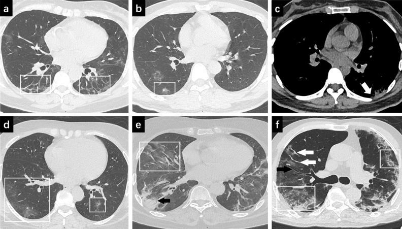
The objective of this study is to expound the CT features of COVID-19 patients whose throat swab samples were negative for two consecutive nucleic acid tests after treatment. We retrospectively reviewed 46 COVID-19 patients with two consecutive negative RT-PCR tests after treatment. The cases were divided into moderate group and severe/critical group according to disease severity. Clinical and CT scanning data were collected. CT signs of pulmonary lesions and the score of lung involvement were expounded. Thirty-nine moderate cases and seven severe/critical cases were included. Residual pulmonary lesions were visible in CT images. Moderate patients showed peripheral lesions while severe/critical cases exhibited both central and peripheral lesions with all lobes involvement. Mixed ground glass opacity (GGO) and pulmonary consolidation were noted. A larger proportion of severe patients showed reticular pulmonary interstitium thickening. Air bronchogram, pleural effusion, vascular enlargement, bronchial wall thickening, bronchiectasis, pleural thickening and pleural adhesion were more frequently observed in severe/critical group. The severe/critical group showed higher CT score. Pulmonary lesions persisted even after twice consecutive negative nucleic acid tests. We strongly recommended regular follow-up of CT scans after nucleic acid tests conversion. Evaluation of complete remission should base on chest CT.

摘要

本研究旨在阐述治疗后连续两次咽拭子核酸检测均为阴性的 COVID-19 患者的 CT 特征。我们回顾性分析了 46 例治疗后连续两次 RT-PCR 检测均为阴性的 COVID-19 患者。根据疾病严重程度将病例分为中度组和重症/危重组。收集临床和 CT 扫描数据。阐述肺部病变的 CT 征象和肺部受累评分。纳入 39 例中度病例和 7 例重症/危重组。CT 图像可见残留肺部病变。中度患者表现为外周病变,而重症/危重组则表现为中央和外周病变,累及所有肺叶。混合磨玻璃密度(GGO)和肺实变。更严重的患者表现出更多的网状肺间质增厚。重症/危重组中更常观察到空气支气管征、胸腔积液、血管增大、支气管壁增厚、支气管扩张、胸膜增厚和胸膜粘连。重症/危重组的 CT 评分更高。即使两次连续核酸检测均为阴性,肺部病变仍持续存在。我们强烈建议在核酸检测转为阴性后定期进行 CT 扫描随访。完全缓解的评估应基于胸部 CT。

https://cdn.ncbi.nlm.nih.gov/pmc/blobs/18a7/7360570/44faa908c066/41598_2020_68509_Fig1_HTML.jpg

相似文献

1
CT features of COVID-19 patients with two consecutive negative RT-PCR tests after treatment.治疗后连续两次 RT-PCR 检测阴性的 COVID-19 患者的 CT 特征。
Sci Rep. 2020 Jul 14;10(1):11548. doi: 10.1038/s41598-020-68509-x.
2
Preliminary CT findings of coronavirus disease 2019 (COVID-19).新型冠状病毒肺炎(COVID-19)的初步 CT 影像学表现。
Clin Imaging. 2020 Sep;65:124-132. doi: 10.1016/j.clinimag.2020.04.042. Epub 2020 May 12.
3
CT imaging features of COVID-19 pneumonia: initial experience from Turkey.土耳其的 COVID-19 肺炎 CT 影像学特征:初步经验。
Diagn Interv Radiol. 2020 Jul;26(4):308-314. doi: 10.5152/dir.2020.20307.
4
CT Features of Coronavirus Disease 2019 (COVID-19) Pneumonia in 62 Patients in Wuhan, China.中国武汉 62 例 2019 年冠状病毒病(COVID-19)肺炎的 CT 特征。
AJR Am J Roentgenol. 2020 Jun;214(6):1287-1294. doi: 10.2214/AJR.20.22975. Epub 2020 Mar 5.
5
A Comparison of Clinical and Chest CT Findings in Patients With Influenza A (H1N1) Virus Infection and Coronavirus Disease (COVID-19).甲型流感病毒(H1N1)感染与新型冠状病毒肺炎(COVID-19)患者的临床和胸部 CT 表现比较。
AJR Am J Roentgenol. 2020 Nov;215(5):1065-1071. doi: 10.2214/AJR.20.23214. Epub 2020 May 26.
6
The chest CT features of coronavirus disease 2019 (COVID-19) in China: a meta-analysis of 19 retrospective studies.中国 2019 年冠状病毒病(COVID-19)的胸部 CT 特征:19 项回顾性研究的荟萃分析。
Virol J. 2020 Oct 21;17(1):159. doi: 10.1186/s12985-020-01432-9.
7
Performance of Radiologists in Differentiating COVID-19 from Non-COVID-19 Viral Pneumonia at Chest CT.放射科医生在胸部 CT 鉴别 COVID-19 与非 COVID-19 病毒性肺炎中的表现。
Radiology. 2020 Aug;296(2):E46-E54. doi: 10.1148/radiol.2020200823. Epub 2020 Mar 10.
8
[Clinical features and high resolution CT imaging evolution of coronavirus disease 2019].新型冠状病毒肺炎的临床特征及高分辨率CT影像学演变
Zhonghua Jie He He Hu Xi Za Zhi. 2020 Jun 12;43(6):509-515. doi: 10.3760/cma.j.cn112147-20200214-00094.
9
Clinical Features and Chest CT Manifestations of Coronavirus Disease 2019 (COVID-19) in a Single-Center Study in Shanghai, China.中国上海单中心研究的 2019 年冠状病毒病(COVID-19)的临床特征和胸部 CT 表现。
AJR Am J Roentgenol. 2020 Jul;215(1):121-126. doi: 10.2214/AJR.20.22959. Epub 2020 Mar 14.
10
Analysis of clinical features and imaging signs of COVID-19 with the assistance of artificial intelligence.人工智能辅助分析 COVID-19 的临床特征和影像征象。
Eur Rev Med Pharmacol Sci. 2020 Aug;24(15):8210-8218. doi: 10.26355/eurrev_202008_22510.

引用本文的文献

1
Serum decorin and biglycan levels as predictive biomarkers for lung fibrosis severity and mortality risk in COVID-19 patients.血清核心蛋白聚糖和双糖链蛋白聚糖水平作为COVID-19患者肺纤维化严重程度和死亡风险的预测生物标志物。
Front Med (Lausanne). 2025 Jan 21;11:1463433. doi: 10.3389/fmed.2024.1463433. eCollection 2024.
2
Machine learning for medical imaging-based COVID-19 detection and diagnosis.用于基于医学影像的新冠病毒检测与诊断的机器学习
Int J Intell Syst. 2021 Sep;36(9):5085-5115. doi: 10.1002/int.22504. Epub 2021 May 31.
3
Unveiling the Clinical Spectrum of Post-COVID-19 Conditions: Assessment and Recommended Strategies.

本文引用的文献

1
Clinical and CT imaging features of 2019 novel coronavirus disease (COVID-19).2019新型冠状病毒病(COVID-19)的临床及CT影像学特征
J Infect. 2020 Jul;81(1):147-178. doi: 10.1016/j.jinf.2020.03.033. Epub 2020 Apr 8.
2
Initial clinical features of suspected coronavirus disease 2019 in two emergency departments outside of Hubei, China.中国湖北省外两家急诊科的 2019 年疑似冠状病毒病的初始临床特征。
J Med Virol. 2020 Sep;92(9):1525-1532. doi: 10.1002/jmv.25763. Epub 2020 Mar 24.
3
Performance of Radiologists in Differentiating COVID-19 from Non-COVID-19 Viral Pneumonia at Chest CT.
揭示新冠后状况的临床谱:评估与推荐策略
Cureus. 2024 Jan 23;16(1):e52827. doi: 10.7759/cureus.52827. eCollection 2024 Jan.
4
Molecular mechanisms of COVID-19-induced pulmonary fibrosis and epithelial-mesenchymal transition.新冠病毒诱导肺纤维化及上皮-间质转化的分子机制
Front Pharmacol. 2023 Aug 3;14:1218059. doi: 10.3389/fphar.2023.1218059. eCollection 2023.
5
Long COVID: a narrative review of the clinical aftermaths of COVID-19 with a focus on the putative pathophysiology and aspects of physical activity.长新冠:对新冠病毒感染临床后果的叙述性综述,重点关注假定的病理生理学和身体活动方面。
Oxf Open Immunol. 2022 Sep 16;3(1):iqac006. doi: 10.1093/oxfimm/iqac006. eCollection 2022.
6
Urokinase System in Pathogenesis of Pulmonary Fibrosis: A Hidden Threat of COVID-19.尿激酶系统在肺纤维化发病机制中的作用:COVID-19 的一个隐藏威胁。
Int J Mol Sci. 2023 Jan 10;24(2):1382. doi: 10.3390/ijms24021382.
7
A scoping review of regulatory T cell dynamics in convalescent COVID-19 patients - indications for their potential involvement in the development of Long COVID?恢复期 COVID-19 患者调节性 T 细胞动态的范围综述——提示其可能参与长新冠的发生?
Front Immunol. 2022 Dec 13;13:1070994. doi: 10.3389/fimmu.2022.1070994. eCollection 2022.
8
Case Report: Pirfenidone in the Treatment of Post-COVID-19 Pulmonary Fibrosis.病例报告:吡非尼酮治疗新冠后肺纤维化
Front Med (Lausanne). 2022 Jun 6;9:925703. doi: 10.3389/fmed.2022.925703. eCollection 2022.
9
Assessing radiomics feature stability with simulated CT acquisitions.评估基于模拟 CT 扫描的放射组学特征稳定性。
Sci Rep. 2022 Mar 18;12(1):4732. doi: 10.1038/s41598-022-08301-1.
10
Role of antifibrotic drugs in the management of post-COVID-19 interstitial lung disease: A review of literature and report from an expert working group.抗纤维化药物在新冠后间质性肺病管理中的作用:文献综述及专家工作组报告
Lung India. 2022 Mar-Apr;39(2):177-186. doi: 10.4103/lungindia.lungindia_659_21.
放射科医生在胸部 CT 鉴别 COVID-19 与非 COVID-19 病毒性肺炎中的表现。
Radiology. 2020 Aug;296(2):E46-E54. doi: 10.1148/radiol.2020200823. Epub 2020 Mar 10.
4
CT Features of Coronavirus Disease 2019 (COVID-19) Pneumonia in 62 Patients in Wuhan, China.中国武汉 62 例 2019 年冠状病毒病(COVID-19)肺炎的 CT 特征。
AJR Am J Roentgenol. 2020 Jun;214(6):1287-1294. doi: 10.2214/AJR.20.22975. Epub 2020 Mar 5.
5
Relation Between Chest CT Findings and Clinical Conditions of Coronavirus Disease (COVID-19) Pneumonia: A Multicenter Study.新型冠状病毒肺炎的胸部 CT 表现与临床特征的关系:一项多中心研究。
AJR Am J Roentgenol. 2020 May;214(5):1072-1077. doi: 10.2214/AJR.20.22976. Epub 2020 Mar 3.
6
Understanding SARS-CoV-2-Mediated Inflammatory Responses: From Mechanisms to Potential Therapeutic Tools.理解 SARS-CoV-2 介导的炎症反应:从机制到潜在的治疗工具。
Virol Sin. 2020 Jun;35(3):266-271. doi: 10.1007/s12250-020-00207-4. Epub 2020 Mar 3.
7
Clinical characteristics and imaging manifestations of the 2019 novel coronavirus disease (COVID-19):A multi-center study in Wenzhou city, Zhejiang, China.中国浙江省温州市多中心研究:2019 年新型冠状病毒病(COVID-19)的临床特征和影像学表现。
J Infect. 2020 Apr;80(4):388-393. doi: 10.1016/j.jinf.2020.02.016. Epub 2020 Feb 26.
8
Chest CT Findings in Patients With Coronavirus Disease 2019 and Its Relationship With Clinical Features.新型冠状病毒肺炎患者的胸部 CT 表现及其与临床特征的关系。
Invest Radiol. 2020 May;55(5):257-261. doi: 10.1097/RLI.0000000000000670.
9
Chest CT Findings in Coronavirus Disease-19 (COVID-19): Relationship to Duration of Infection.胸部计算机断层扫描在 2019 年冠状病毒病(COVID-19)中的表现:与感染持续时间的关系。
Radiology. 2020 Jun;295(3):200463. doi: 10.1148/radiol.2020200463. Epub 2020 Feb 20.
10
CT Imaging Features of 2019 Novel Coronavirus (2019-nCoV).2019 新型冠状病毒(2019-nCoV)的 CT 影像学特征。
Radiology. 2020 Apr;295(1):202-207. doi: 10.1148/radiol.2020200230. Epub 2020 Feb 4.