Computer Vision Lab, ETH Zurich, Zurich, Switzerland.
University of Applied Sciences Western Switzerland (HES-SO) Valais, Sierre, Switzerland.
Sci Rep. 2022 Mar 18;12(1):4732. doi: 10.1038/s41598-022-08301-1.
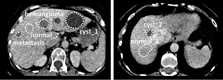
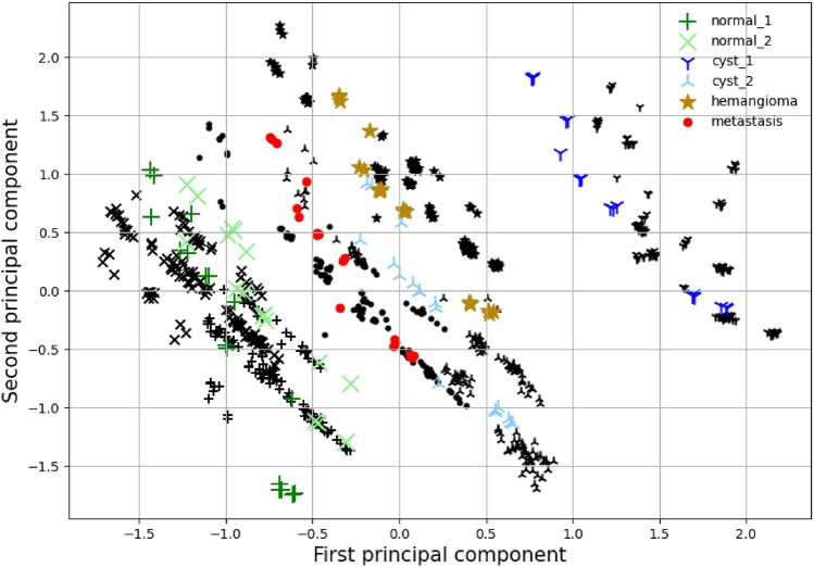
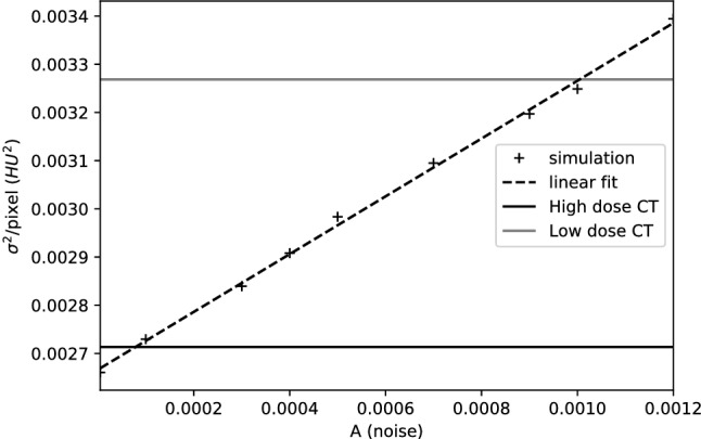
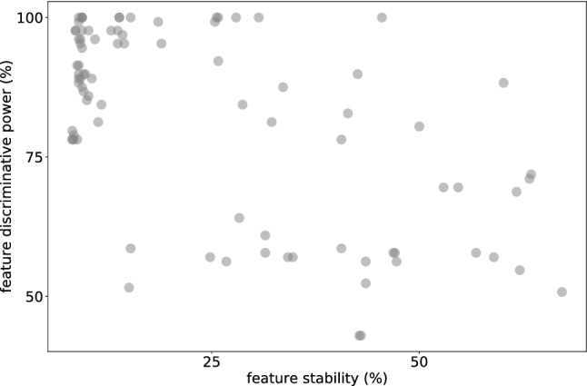
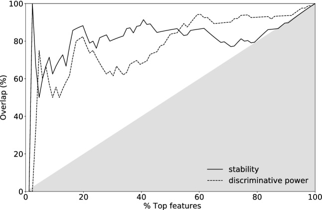
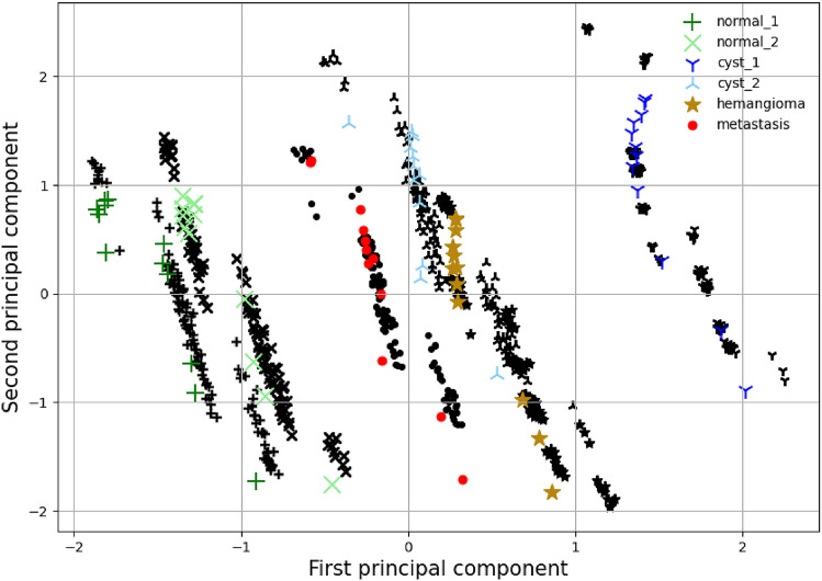
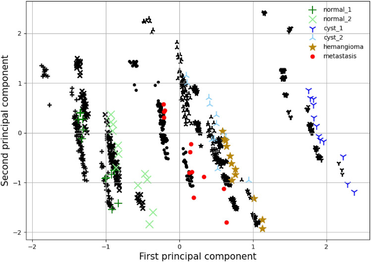
Medical imaging quantitative features had once disputable usefulness in clinical studies. Nowadays, advancements in analysis techniques, for instance through machine learning, have enabled quantitative features to be progressively useful in diagnosis and research. Tissue characterisation is improved via the "radiomics" features, whose extraction can be automated. Despite the advances, stability of quantitative features remains an important open problem. As features can be highly sensitive to variations of acquisition details, it is not trivial to quantify stability and efficiently select stable features. In this work, we develop and validate a Computed Tomography (CT) simulator environment based on the publicly available ASTRA toolbox ( www.astra-toolbox.com ). We show that the variability, stability and discriminative power of the radiomics features extracted from the virtual phantom images generated by the simulator are similar to those observed in a tandem phantom study. Additionally, we show that the variability is matched between a multi-center phantom study and simulated results. Consequently, we demonstrate that the simulator can be utilised to assess radiomics features' stability and discriminative power.
医学影像定量特征在临床研究中曾经具有争议性的有用性。如今,分析技术的进步,例如通过机器学习,已经使得定量特征在诊断和研究中逐渐变得有用。通过“放射组学”特征可以改善组织特征描述,其提取可以自动化。尽管取得了这些进展,但定量特征的稳定性仍然是一个重要的开放性问题。由于特征可能对采集细节的变化非常敏感,因此量化稳定性并有效地选择稳定特征并非易事。在这项工作中,我们基于公开可用的 ASTRA 工具箱(www.astra-toolbox.com)开发并验证了一种计算机断层扫描(CT)模拟器环境。我们表明,从模拟器生成的虚拟体模图像中提取的放射组学特征的可变性、稳定性和区分能力与在串联体模研究中观察到的特征相似。此外,我们表明,多中心体模研究和模拟结果之间的可变性相匹配。因此,我们证明了模拟器可用于评估放射组学特征的稳定性和区分能力。