Wang Yamin, Zhang Min, Sun Ying, Wang Xiaohui, Song Zhaowei, Li Huazhang, Liu Kexin, Li Zhijian
Department of Ophthalmology, the First Affiliated Hospital, Harbin Medical University, 143 Yiman Street, Nangang District, Harbin, China.
Department of Ophthalmology, the 2nd Hospital of Heilongjiang, Harbin, China.
BMC Ophthalmol. 2020 Jul 15;20(1):289. doi: 10.1186/s12886-020-01565-z.
With the popularity of blue-rich light-emitting diode (LED)-backlit display devices, our eyes are now exposed to more short-wavelength blue light than they were in the past. The goal of this study was to investigate the pathogenesis of cataracts after short-wavelength light exposure.
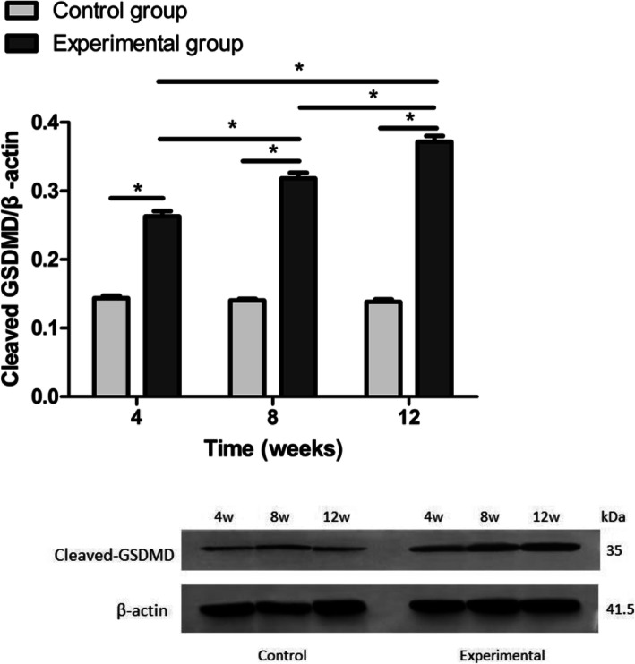
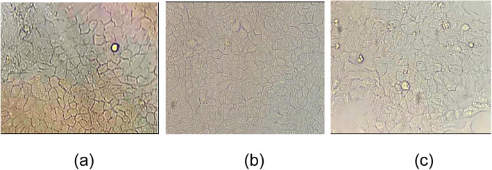
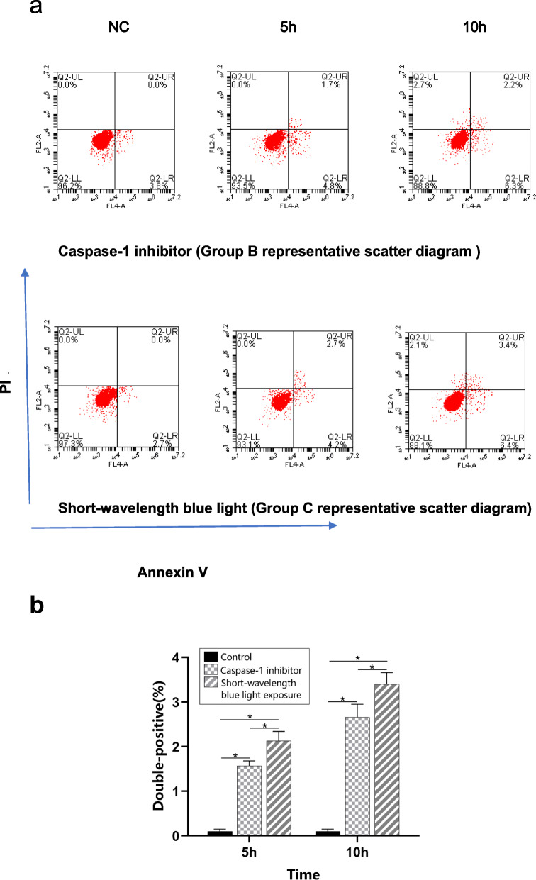
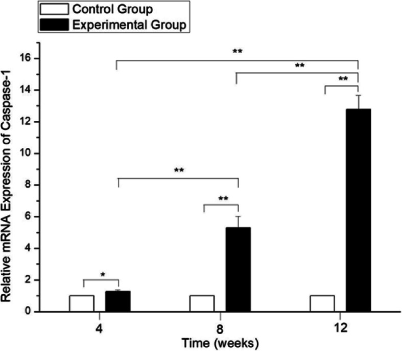
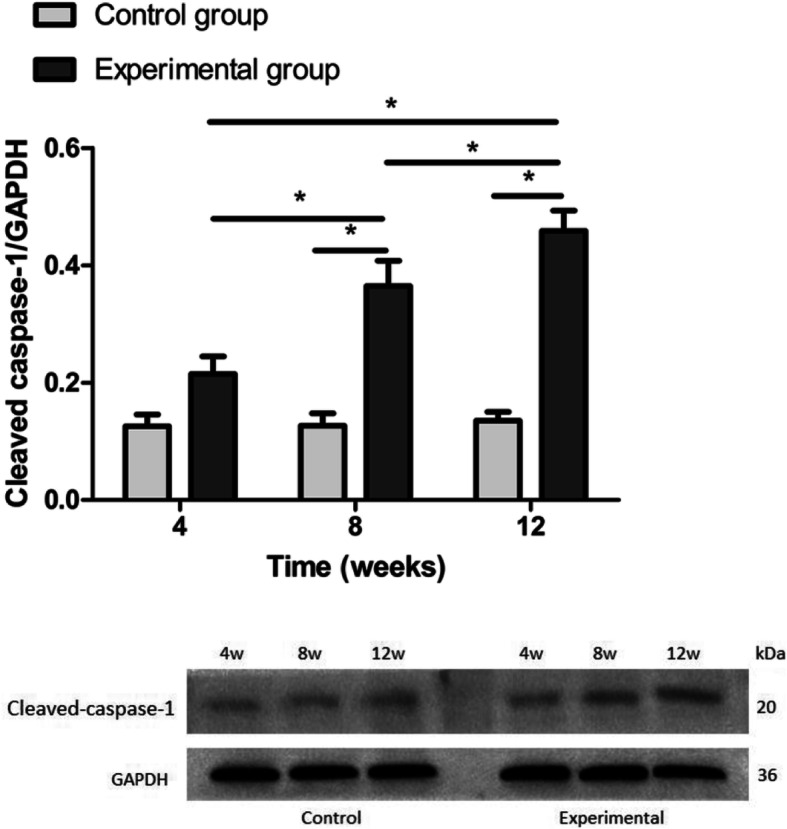
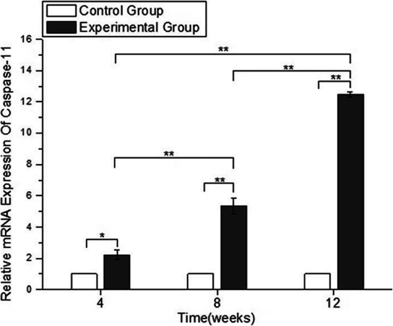
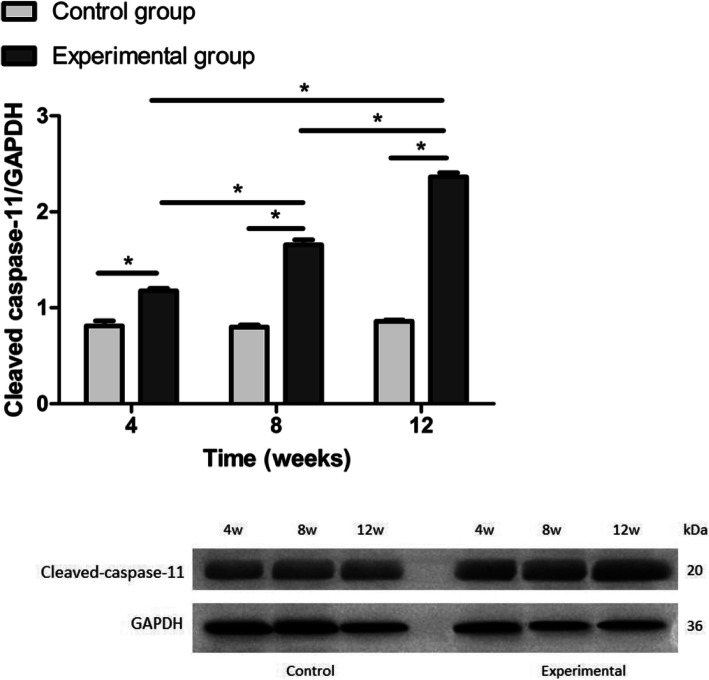
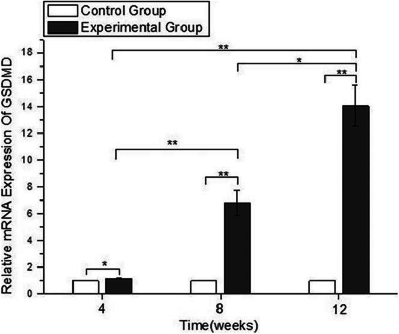
Sprague-Dawley (SD) rats were selected and randomly divided into a control group (10 rats each for the 4-, 8-, and 12-week groups) and an experimental group (10 rats each for the 4-, 8-, and 12-week groups). The rats in the experimental group were exposed to a short-wavelength blue LED lamp for 12 h per day. After exposure to the blue LED lamp, the rats were maintained in total darkness for 12 h, after which a 12-h light/dark cycle was resumed. The intensity of the lamp was 3000 lx. At the end of the short-wavelength blue LED lamp exposure (for 4, 8, and 12 weeks), the expression levels of caspase-1, caspase-11 and gasdermin D (GSDMD) were examined in rat lens epithelial cells (LECs) using qRT-PCR and Western blot analyses. An illuminance of 2500 lx was used to study the potential effect of blue LED light on HLE-B3 hLECs in vitro. AC-YVAD-CMK, a caspase-1 inhibitor, was used to confirm the pyroptosis of LECs by flow cytometry.
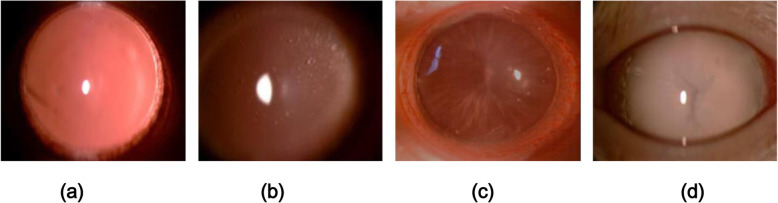
After 6 weeks, cataracts developed in the experimental rats (4/20 eyes). The clarity of the lens gradually worsened with the duration of exposure. Twelve weeks later, all of the rat eyes had developed cataracts. The expression levels of caspase-1, caspase-11 and GSDMD at 4, 8, and 12 weeks were significantly higher in the samples from rats exposed to a short-wavelength blue LED lamp than in the samples from control rats (p<0.05). The proportions of double-positive hLECs were significantly increased in the 5-h and 10-h short-wavelength blue light exposure subgroups compared with the 5-h and 10-h caspase-1 inhibitor subgroups (p < 0.05).
The data indicate that pyroptosis plays a key role in cataract induction after short-wavelength blue light exposure. This study might provide new insights into a novel pathogenic mechanism of cataracts.
随着富含蓝光的发光二极管(LED)背光显示设备的普及,我们的眼睛现在比过去接触到更多的短波长蓝光。本研究的目的是探讨短波长光暴露后白内障的发病机制。
选用Sprague-Dawley(SD)大鼠,随机分为对照组(4周、8周和12周组每组10只大鼠)和实验组(4周、8周和12周组每组10只大鼠)。实验组大鼠每天暴露于短波长蓝色LED灯下12小时。暴露于蓝色LED灯后,大鼠在完全黑暗中饲养12小时,之后恢复12小时光照/黑暗循环。灯的强度为3000勒克斯。在短波长蓝色LED灯暴露结束时(4周、8周和12周),使用qRT-PCR和蛋白质印迹分析检测大鼠晶状体上皮细胞(LECs)中caspase-1、caspase-11和gasdermin D(GSDMD)的表达水平。使用2500勒克斯的照度研究蓝色LED光对体外培养的人晶状体上皮细胞(HLE-B3 hLECs)的潜在影响。使用caspase-1抑制剂AC-YVAD-CMK通过流式细胞术确认LECs的焦亡。
6周后实验组大鼠出现白内障(4/20只眼)。晶状体的清晰度随着暴露时间的延长而逐渐恶化。12周后,所有大鼠眼睛均出现白内障。暴露于短波长蓝色LED灯的大鼠样本中,4周、8周和12周时caspase-11、caspase-11和GSDMD的表达水平显著高于对照组大鼠样本(p<0.05)。与5小时和10小时caspase-1抑制剂亚组相比,5小时和10小时短波长蓝光暴露亚组中双阳性hLECs的比例显著增加(p<0.05)。
数据表明,焦亡在短波长蓝光暴露后诱导白内障中起关键作用。本研究可能为白内障的新型致病机制提供新的见解。