Li Fengwu, Geng Xiaokun, Huber Christian, Stone Christopher, Ding Yuchuan
China-America Institute of Neuroscience, Luhe Hospital, Capital Medical University, Beijing, China.
Department of Neurology, Beijing Luhe Hospital, Capital Medical University, Beijing, China.
Front Cell Neurosci. 2020 Jun 25;14:186. doi: 10.3389/fncel.2020.00186. eCollection 2020.
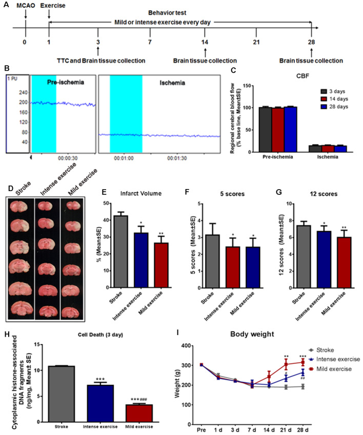
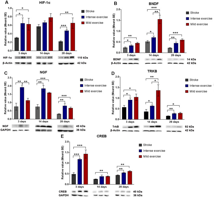
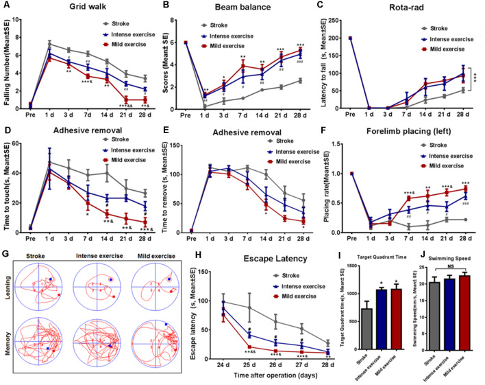
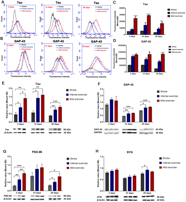
Although physical exercise has been demonstrated to augment recovery of the post-stroke brain, the question of what level of exercise intensity optimizes neurological outcomes of post-stroke rehabilitation remains unsettled. In this study, we aim to clarify the mechanisms underlying the intensity-dependent effect of exercise on neurologic function, and thereby to help direct the clinical application of exercise-based neurorehabilitation. To do this, we used a well-established rat model of ischemic stroke consisting of cerebral ischemia induction through middle cerebral artery occlusion (MCAO). Ischemic rats were subsequently assigned either to a control group entailing post-stroke rest or to one of two exercise groups distinguished by the intensity of their accompanying treadmill regimens. After 24 h of reperfusion, exercise was initiated. Infarct volume, apoptotic cell death, and neurological defects were quantified in all groups at 3 days, and motor and cognitive functions were tracked up to day-28. Additionally, Western blotting was used to assess the influence of our interventions on several proteins related to synaptogenesis and neuroplasticity (growth-associated protein 43, a microtubule-associated protein, postsynaptic density-95, synapsin I, hypoxia-inducible factor-1α, brain-derived neurotrophic factor, nerve growth factor, tyrosine kinase B, and cAMP response element-binding protein). Our results were in equal parts encouraging and surprising. Both mild and intense exercise significantly decreased infarct volume, cell death, and neurological deficits. Motor and cognitive function, as determined using an array of tests such as beam balance, forelimb placing, and the Morris water maze, were also significantly improved by both exercise protocols. Interestingly, while an obvious enhancement of neuroplasticity proteins was shown in both exercise groups, mild exercise rats demonstrated a stronger effect on the expressions of Tau ( < 0.01), brain-derived neurotrophic factor ( < 0.01), and tyrosine kinase B ( < 0.05). These findings contribute to the growing body of literature regarding the positive effects of both mild and intense long-term treadmill exercise on brain injury, functional outcome, and neuroplasticity. Additionally, the results may provide a base for our future study regarding the regulation of HIF-1α on the BDNF/TrkB/CREB pathway in the biochemical processes underlying post-stroke synaptic plasticity.
尽管体育锻炼已被证明可促进中风后脑的恢复,但何种运动强度能使中风后康复的神经学结果达到最佳这一问题仍未得到解决。在本研究中,我们旨在阐明运动对神经功能的强度依赖性效应背后的机制,从而有助于指导基于运动的神经康复的临床应用。为此,我们使用了一种成熟的缺血性中风大鼠模型,该模型通过大脑中动脉闭塞(MCAO)诱导脑缺血。随后,将缺血大鼠分为对照组(中风后休息)或两个运动组之一,这两个运动组根据其伴随的跑步机训练强度进行区分。再灌注24小时后开始运动。在第3天对所有组的梗死体积、凋亡细胞死亡和神经功能缺损进行量化,并追踪至第28天的运动和认知功能。此外,采用蛋白质免疫印迹法评估我们的干预措施对几种与突触形成和神经可塑性相关蛋白质的影响(生长相关蛋白43、一种微管相关蛋白、突触后密度蛋白95、突触素I、缺氧诱导因子-1α、脑源性神经营养因子、神经生长因子、酪氨酸激酶B和环磷酸腺苷反应元件结合蛋白)。我们的结果既令人鼓舞又令人惊讶。轻度和高强度运动均显著降低了梗死体积、细胞死亡和神经功能缺损。通过一系列测试(如光束平衡、前肢放置和莫里斯水迷宫)确定的运动和认知功能,在两种运动方案中也均得到显著改善。有趣的是,虽然两个运动组均显示神经可塑性蛋白明显增强,但轻度运动大鼠对Tau(<0.01)、脑源性神经营养因子(<0.01)和酪氨酸激酶B(<0.05)的表达有更强的影响。这些发现为越来越多关于轻度和高强度长期跑步机运动对脑损伤、功能结果和神经可塑性的积极影响的文献做出了贡献。此外,这些结果可能为我们未来关于缺氧诱导因子-1α在中风后突触可塑性生化过程中对脑源性神经营养因子/酪氨酸激酶B/环磷酸腺苷反应元件结合蛋白途径调节的研究提供基础。