Sun Jintao, Gao Yuan, Miao Ailiang, Yu Chuanyong, Tang Lu, Huang Shuyang, Wu Caiyun, Shi Qi, Zhang Tingting, Li Yihan, Sun Yulei, Wang Xiaoshan
Department of Neurology, The Affiliated Brain Hospital of Nanjing Medical University, Nanjing Medical University, Nanjing, China.
Front Hum Neurosci. 2020 Jun 26;14:221. doi: 10.3389/fnhum.2020.00221. eCollection 2020.
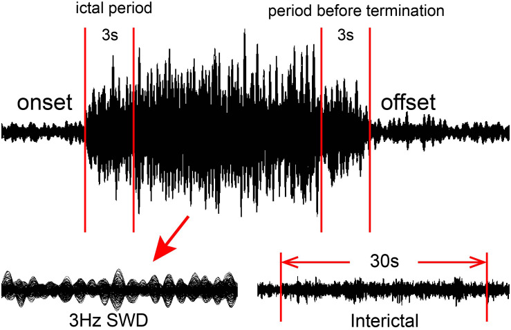
This study aimed to investigate the spectral and spatial signatures of neuromagnetic activity underlying the termination of absence seizures.
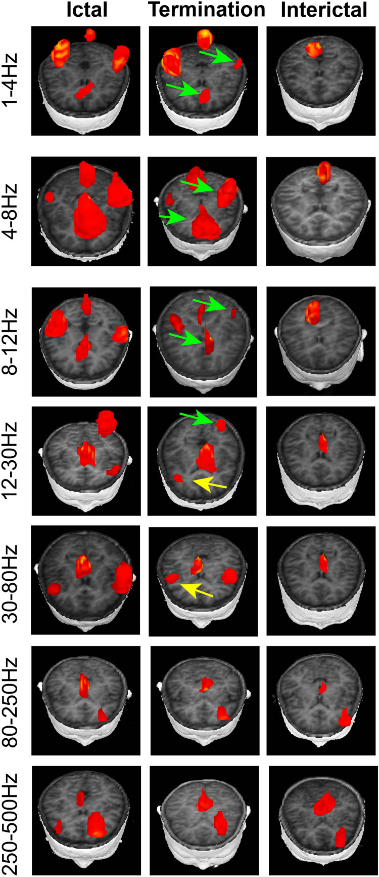
Magnetoencephalography (MEG) data were recorded from 18 drug-naive patients with childhood absence epilepsy (CAE). Accumulated source imaging (ASI) was used to analyze MEG data at the source level in seven frequency ranges: delta (1-4 Hz), theta (4-8 Hz), alpha (8-12 Hz), beta (12-30 Hz), gamma (30-80 Hz), ripple (80-250 Hz), and fast ripple (250-500 Hz).
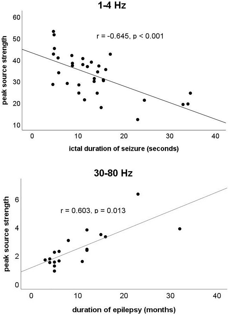
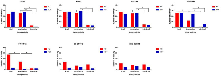
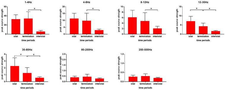
In the 1-4, 4-8, and 8-12 Hz ranges, the magnetic source during seizure termination appeared to be consistent over the ictal period and was mainly localized in the frontal cortex (FC) and parieto-occipito-temporal junction (POT). In the 12-30 and 30-80 Hz ranges, a significant reduction in source activity was observed in the frontal lobe during seizure termination as well as a decrease in peak source strength. The ictal peak source strength in the 1-4 Hz range was negatively correlated with the ictal duration of the seizure, whereas in the 30-80 Hz range, it was positively correlated with the course of epilepsy.
The termination of absence seizures is associated with a dynamic neuromagnetic process. Frequency-dependent changes in the FC were observed during seizure termination, which may be involved in the process of neural network interaction. Neuromagnetic activity in different frequency bands may play different roles in the pathophysiological mechanism during absence seizures.
本研究旨在探究失神发作终止时神经磁活动的频谱和空间特征。
记录了18例未经药物治疗的儿童失神癫痫(CAE)患者的脑磁图(MEG)数据。采用累积源成像(ASI)在七个频率范围对MEG数据进行源水平分析:δ(1 - 4赫兹)、θ(4 - 8赫兹)、α(8 - 12赫兹)、β(12 - 30赫兹)、γ(30 - 80赫兹)、涟漪波(80 - 250赫兹)和快涟漪波(250 - 500赫兹)。
在1 - 4赫兹、4 - 8赫兹和8 - 12赫兹范围内,发作终止期间的磁源在发作期似乎是一致的,主要定位于额叶皮质(FC)和顶枕颞交界处(POT)。在12 - 30赫兹和30 - 80赫兹范围内,发作终止期间额叶的源活动显著降低,同时源峰值强度也降低。1 - 4赫兹范围内的发作期源峰值强度与发作的发作持续时间呈负相关,而在30 - 80赫兹范围内,它与癫痫病程呈正相关。
失神发作的终止与动态神经磁过程相关。发作终止期间观察到额叶皮质存在频率依赖性变化,这可能参与神经网络相互作用过程。不同频段的神经磁活动在失神发作的病理生理机制中可能发挥不同作用。