• 文献检索
  • 文档翻译
  • 深度研究
  • 学术资讯
  • Suppr Zotero 插件Zotero 插件
  • 邀请有礼
  • 套餐&价格
  • 历史记录
应用&插件
Suppr Zotero 插件Zotero 插件浏览器插件Mac 客户端Windows 客户端微信小程序
定价
高级版会员购买积分包购买API积分包
服务
文献检索文档翻译深度研究API 文档MCP 服务
关于我们
关于 Suppr公司介绍联系我们用户协议隐私条款
关注我们

Suppr 超能文献

核心技术专利:CN118964589B侵权必究
粤ICP备2023148730 号-1Suppr @ 2026

文献检索

告别复杂PubMed语法,用中文像聊天一样搜索,搜遍4000万医学文献。AI智能推荐,让科研检索更轻松。

立即免费搜索

文件翻译

保留排版,准确专业,支持PDF/Word/PPT等文件格式,支持 12+语言互译。

免费翻译文档

深度研究

AI帮你快速写综述,25分钟生成高质量综述,智能提取关键信息,辅助科研写作。

立即免费体验

心脏磁共振成像通过间接方法系统性地高估二尖瓣反流情况。

Cardiac magnetic resonance systematically overestimates mitral regurgitations by the indirect method.

作者信息

Bertelsen Litten, Vejlstrup Niels, Andreasen Laura, Olesen Morten Salling, Svendsen Jesper Hastrup

机构信息

Department of Cardiology, Centre for Cardiac, Vascular, Pulmonary and Infectious Diseases, Rigshospitalet, Copenhagen University Hospital, Copenhagen, Denmark

Department of Cardiology, Centre for Cardiac, Vascular, Pulmonary and Infectious Diseases, Rigshospitalet, Copenhagen University Hospital, Copenhagen, Denmark.

出版信息

Open Heart. 2020 Jul;7(2). doi: 10.1136/openhrt-2020-001323.

DOI:10.1136/openhrt-2020-001323
PMID:32675299
原文链接:https://pmc.ncbi.nlm.nih.gov/articles/PMC7368492/
Abstract

OBJECTIVE

Cardiac MRI is quickly emerging as the gold standard for assessment of mitral regurgitation, most commonly with the indirect method subtracting forward flow in aorta from volumetric segmentation of the left ventricle. We aimed to investigate how aortic flow measurements with increasing distance from the aortic valve affect calculated mitral regurgitations and whether measurements were influenced by breath-hold regimen.

METHODS

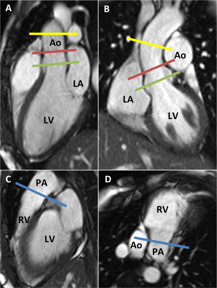
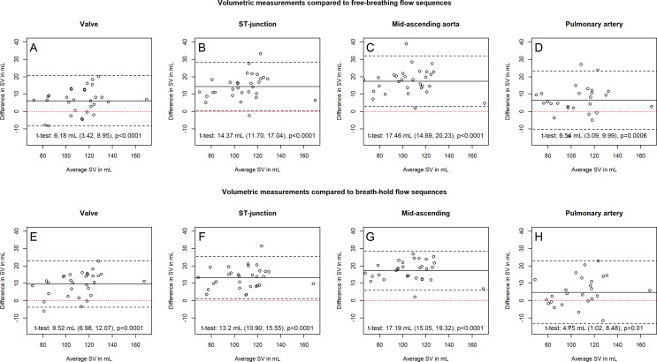
Free-breathing and breath-hold phase contrast flows were measured in aorta at valve level, sinotubular (ST) junction, mid-ascending aorta and in the pulmonary trunk. Flow measurements were pairwise compared, and subsequently, after exclusion of patients with visible mitral and tricuspid regurgitations for left-sided and right-sided comparisons, respectively, flow-measured stroke volumes were compared with ventricular volumetric segmentations.

RESULTS

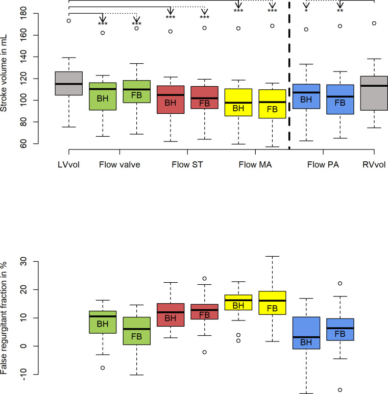
Thirty-nine participants without arrhythmias or structural abnormalities of the large vessels were included. Stroke volumes measured with free-breathing and breath-hold flow decreased equally with increasing distance to the aortic valves (breath-hold flow: aortic valve 105.6±20.8 mL, ST junction 101.5±20.7 mL, mid-ascending aorta 98.1±21.5 mL). After exclusion of atrioventricular regurgitations, stroke volumes determined by volumetric measurements were higher compared with values determined by flow measurements, corresponding to 'false' atrioventricular regurgitations of 8.0%±5.8% with flow measured at valve level, 11.6%±5.2% at the ST junction and 15.3%±5.0% at the mid-ascending aorta.

CONCLUSIONS

Stroke volumes determined by flow decrease throughout the proximal aorta and are systematically lower than volumetrically measured stroke volumes. The indirect method systematically overestimates mitral regurgitations, especially with increasing distance from the aortic valves.

摘要

目的

心脏磁共振成像正迅速成为评估二尖瓣反流的金标准,最常用的间接方法是通过左心室容积分割减去主动脉内的前向血流。我们旨在研究距主动脉瓣距离增加时主动脉血流测量如何影响计算出的二尖瓣反流,以及测量是否受屏气方案的影响。

方法

在主动脉瓣水平、窦管(ST)交界处、升主动脉中部和肺动脉主干处,对自由呼吸和屏气状态下的相位对比血流进行测量。对血流测量值进行两两比较,随后,在分别排除左侧和右侧可见二尖瓣和三尖瓣反流的患者后,将血流测量的每搏输出量与心室容积分割结果进行比较。

结果

纳入39名无心律失常或大血管结构异常的参与者。自由呼吸和屏气血流测量的每搏输出量均随距主动脉瓣距离增加而同等程度下降(屏气血流:主动脉瓣处105.6±20.8 mL,ST交界处101.5±20.7 mL,升主动脉中部98.1±21.5 mL)。排除房室反流后,容积测量确定的每搏输出量高于血流测量值,对应于在瓣膜水平血流测量时“假性”房室反流为8.0%±5.8%,在ST交界处为11.6%±5.2%,在升主动脉中部为15.3%±5.0%。

结论

通过血流测定的每搏输出量在整个近端主动脉中均下降,且系统性低于容积测量的每搏输出量。间接方法系统性高估二尖瓣反流,尤其是距主动脉瓣距离增加时。

https://cdn.ncbi.nlm.nih.gov/pmc/blobs/1cf3/7368492/5afdd5f658b4/openhrt-2020-001323f04.jpg
https://cdn.ncbi.nlm.nih.gov/pmc/blobs/1cf3/7368492/cab3aeebbfb9/openhrt-2020-001323f01.jpg
https://cdn.ncbi.nlm.nih.gov/pmc/blobs/1cf3/7368492/4291886e8a5b/openhrt-2020-001323f02.jpg
https://cdn.ncbi.nlm.nih.gov/pmc/blobs/1cf3/7368492/fcf7db8172b5/openhrt-2020-001323f03.jpg
https://cdn.ncbi.nlm.nih.gov/pmc/blobs/1cf3/7368492/5afdd5f658b4/openhrt-2020-001323f04.jpg

相似文献

1
Cardiac magnetic resonance systematically overestimates mitral regurgitations by the indirect method.心脏磁共振成像通过间接方法系统性地高估二尖瓣反流情况。
Open Heart. 2020 Jul;7(2). doi: 10.1136/openhrt-2020-001323.
2
Flow measurement at the aortic root - impact of location of through-plane phase contrast velocity mapping.主动脉根部血流测量——平面内相位对比速度映射位置的影响
J Cardiovasc Magn Reson. 2016 Sep 7;18(1):55. doi: 10.1186/s12968-016-0277-7.
3
Quantification of mitral regurgitation in patients with hypertrophic cardiomyopathy using aortic and pulmonary flow data: impacts of left ventricular outflow tract obstruction and different left ventricular segmentation methods.应用主动脉和肺血流数据对肥厚型心肌病患者二尖瓣反流进行定量评估:左心室流出道梗阻和不同左心室分段方法的影响。
J Cardiovasc Magn Reson. 2017 Dec 21;19(1):105. doi: 10.1186/s12968-017-0417-8.
4
Effect of protocol choice on phase contrast cardiac magnetic resonance flow measurement in the ascending aorta: breath-hold and non-breath-hold.协议选择对升主动脉相位对比心脏磁共振流量测量的影响:屏气和非屏气。
Int J Cardiovasc Imaging. 2013 Jan;29(1):113-20. doi: 10.1007/s10554-012-0047-z. Epub 2012 Apr 22.
5
[Biventricular MR volumetric analysis and MR flow quantification in the ascending aorta and pulmonary trunk for quantification of valvular regurgitation].[双心室磁共振容积分析及升主动脉和肺动脉干磁共振血流定量用于评估瓣膜反流]
Rofo. 2004 Mar;176(3):342-9. doi: 10.1055/s-2004-812751.
6
Quantitating aortic regurgitation by cardiovascular magnetic resonance: significant variations due to slice location and breath holding.通过心血管磁共振定量主动脉瓣反流:因层面位置和屏气导致的显著差异。
Eur Radiol. 2016 Sep;26(9):3180-9. doi: 10.1007/s00330-015-4120-6. Epub 2015 Dec 3.
7
Assessment of mitral regurgitation by 3-dimensional proximal flow convergence using magnetic resonance imaging: comparison with echo-Doppler.利用磁共振成像通过三维近端血流会聚评估二尖瓣反流:与超声多普勒的比较。
Int J Cardiovasc Imaging. 2018 May;34(5):793-802. doi: 10.1007/s10554-017-1290-0. Epub 2017 Dec 19.
8
Mitral regurgitation quantification by cardiovascular magnetic resonance: a comparison of indirect quantification methods.心血管磁共振对二尖瓣反流的定量分析:间接定量方法的比较
Int J Cardiovasc Imaging. 2015 Aug;31(6):1223-31. doi: 10.1007/s10554-015-0681-3. Epub 2015 May 23.
9
[Comparison between biventricular cine MRI and MR flow quantification in ascending aorta and pulmonary outflow tract for the assessment of intracardial shunt volumes].[双心室电影磁共振成像与升主动脉及肺动脉流出道磁共振血流定量在评估心内分流容积中的比较]
Rofo. 2002 Nov;174(11):1380-6. doi: 10.1055/s-2002-35336.
10
Cardiac remodelling and function with primary mitral valve insufficiency studied by magnetic resonance imaging.通过磁共振成像研究原发性二尖瓣关闭不全时的心脏重塑与功能
Eur Heart J Cardiovasc Imaging. 2016 Aug;17(8):863-70. doi: 10.1093/ehjci/jev321. Epub 2016 Jan 12.

引用本文的文献

1
Tricuspid valve flow measurement using a deep learning framework for automated valve-tracking 2D phase contrast.使用深度学习框架进行自动化瓣跟踪二维相位对比的三尖瓣流量测量。
Magn Reson Med. 2024 Nov;92(5):1838-1850. doi: 10.1002/mrm.30163. Epub 2024 May 31.
2
Comparative Study of 2D-Cine and 3D-wh Volumetry: Revealing Systemic Error of 2D-Cine Volumetry.二维电影成像与三维全容积成像的比较研究:揭示二维电影成像容积测量的系统误差
Diagnostics (Basel). 2023 Oct 10;13(20):3162. doi: 10.3390/diagnostics13203162.
3
Evaluation of left ventricular and left atrial volumetric function from native MR multislice 4D flow magnitude data.

本文引用的文献

1
Assessment of mitral valve regurgitation by cardiovascular magnetic resonance imaging.心血管磁共振成像评估二尖瓣反流。
Nat Rev Cardiol. 2020 May;17(5):298-312. doi: 10.1038/s41569-019-0305-z. Epub 2019 Dec 9.
2
CMR quantitation of change in mitral regurgitation following transcatheter aortic valve replacement (TAVR): impact on left ventricular reverse remodeling and outcome.经导管主动脉瓣置换术(TAVR)后二尖瓣反流变化的心脏磁共振成像(CMR)定量分析:对左心室逆向重构及预后的影响
Int J Cardiovasc Imaging. 2019 Jan;35(1):161-170. doi: 10.1007/s10554-018-1441-y. Epub 2018 Sep 4.
3
Use of Cardiac Magnetic Resonance Imaging in Assessing Mitral Regurgitation: Current Evidence.
从原生 MR 多层 4D 流场幅度数据评估左心室和左心房容积功能。
Eur Radiol. 2024 Feb;34(2):981-993. doi: 10.1007/s00330-023-10017-3. Epub 2023 Aug 15.
4
Quantification of primary mitral regurgitation by echocardiography: A practical appraisal.超声心动图对原发性二尖瓣反流的定量评估:实用评价
Front Cardiovasc Med. 2023 Mar 10;10:1107724. doi: 10.3389/fcvm.2023.1107724. eCollection 2023.
5
Role of Cardiovascular Magnetic Resonance in Native Valvular Regurgitation: A Comprehensive Review of Protocols, Grading of Severity, and Prediction of Valve Surgery.心血管磁共振在原发性瓣膜反流中的作用:协议、严重程度分级及瓣膜手术预测的综合综述
Front Cardiovasc Med. 2022 Jul 7;9:881141. doi: 10.3389/fcvm.2022.881141. eCollection 2022.
6
Reduced External Iliac Venous Blood Flow Rate Is Associated with Asymptomatic Compression of the Common Iliac Veins.髂外静脉血流速率降低与无症状性髂总静脉受压相关。
Medicina (Kaunas). 2021 Aug 18;57(8):835. doi: 10.3390/medicina57080835.
使用心脏磁共振成像评估二尖瓣反流:当前证据。
J Am Coll Cardiol. 2018 Feb 6;71(5):547-563. doi: 10.1016/j.jacc.2017.12.009.
4
Cardiovascular magnetic resonance in the evaluation of heart valve disease.心血管磁共振成像在心脏瓣膜病评估中的应用
BMC Med Imaging. 2017 Dec 29;17(1):67. doi: 10.1186/s12880-017-0238-0.
5
Prognostic Implications of Magnetic Resonance-Derived Quantification in Asymptomatic Patients With Organic Mitral Regurgitation: Comparison With Doppler Echocardiography-Derived Integrative Approach.磁共振衍生定量分析在无症状性器质性二尖瓣反流患者中的预后意义:与多普勒超声心动图衍生综合方法的比较。
Circulation. 2018 Mar 27;137(13):1349-1360. doi: 10.1161/CIRCULATIONAHA.117.029332. Epub 2017 Dec 21.
6
Quantification of mitral regurgitation in patients with hypertrophic cardiomyopathy using aortic and pulmonary flow data: impacts of left ventricular outflow tract obstruction and different left ventricular segmentation methods.应用主动脉和肺血流数据对肥厚型心肌病患者二尖瓣反流进行定量评估:左心室流出道梗阻和不同左心室分段方法的影响。
J Cardiovasc Magn Reson. 2017 Dec 21;19(1):105. doi: 10.1186/s12968-017-0417-8.
7
Clinical applications of intra-cardiac four-dimensional flow cardiovascular magnetic resonance: A systematic review.心脏内四维血流心血管磁共振的临床应用:系统评价。
Int J Cardiol. 2017 Dec 15;249:486-493. doi: 10.1016/j.ijcard.2017.07.023. Epub 2017 Sep 28.
8
Are Echocardiography and CMR Really Discordant in Mitral Regurgitation?超声心动图和心脏磁共振成像在二尖瓣反流中真的不一致吗?
JACC Cardiovasc Imaging. 2017 Jul;10(7):823-824. doi: 10.1016/j.jcmg.2016.08.007. Epub 2016 Nov 16.
9
Quantitation of mitral regurgitation with cardiac magnetic resonance imaging: a systematic review.心脏磁共振成像定量评估二尖瓣反流:一项系统评价
Heart. 2016 Dec 1;102(23):1864-1870. doi: 10.1136/heartjnl-2015-309054. Epub 2016 Oct 12.
10
Effects of slice orientation on reproducibility of sequential assessment of right ventricular volumes and ejection fraction: short-axis vs transverse SSFP cine cardiovascular magnetic resonance.层面取向对右心室容积及射血分数连续评估可重复性的影响:短轴与横轴稳态自由感应衰减电影心血管磁共振成像对比
J Cardiovasc Magn Reson. 2016 Sep 22;18(1):60. doi: 10.1186/s12968-016-0282-x.