Department of Energy and Materials Engineering, Dongguk University-Seoul, 30 Pildong-ro 1-gil, Seoul 04620, Republic of Korea.
Department of Biological Engineering, Biohybrid Systems Research Center (BSRC), Inha University, 100 Inha-ro, Nam-gu, Incheon 22212, Republic of Korea.
Theranostics. 2020 Jun 22;10(17):7841-7856. doi: 10.7150/thno.42291. eCollection 2020.
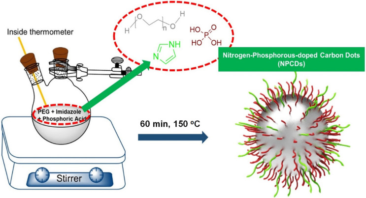
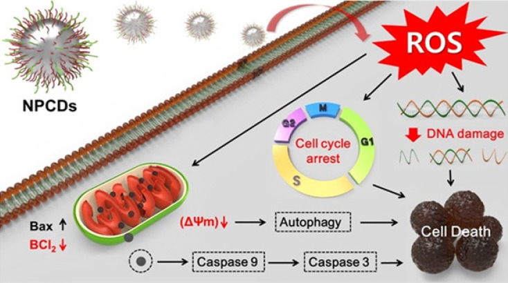
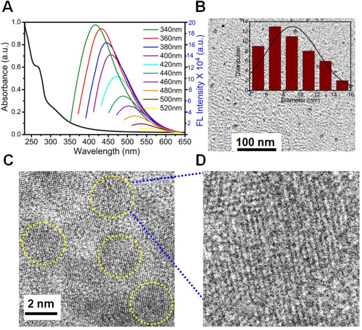
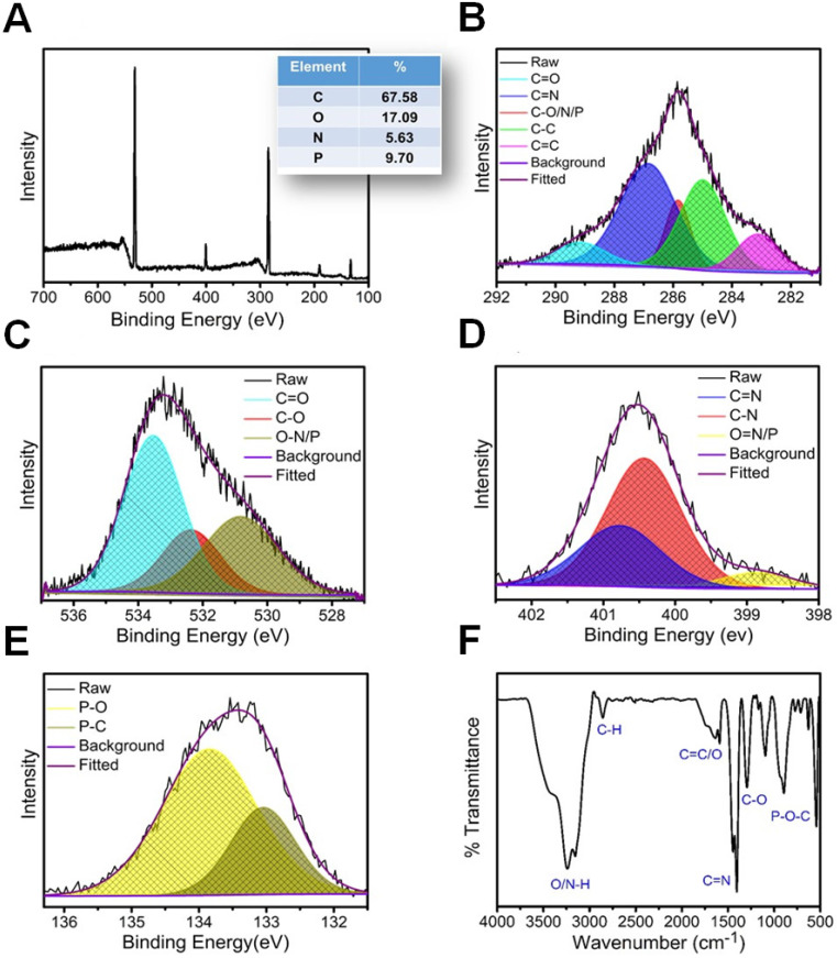
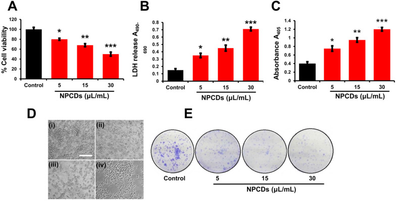
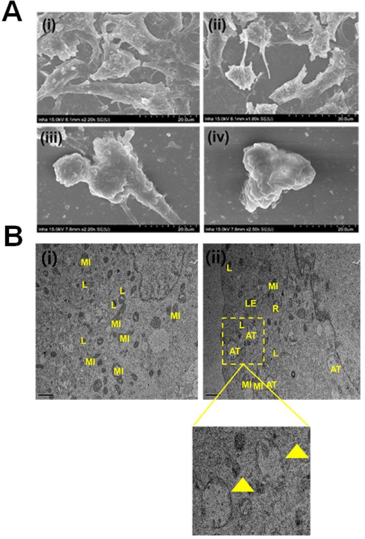
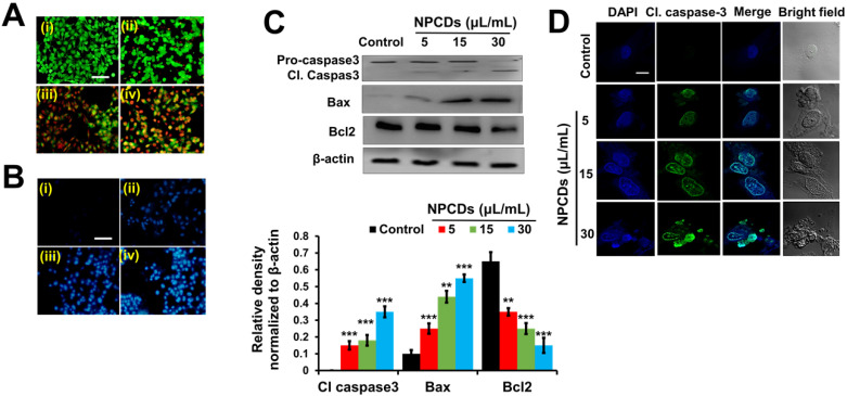
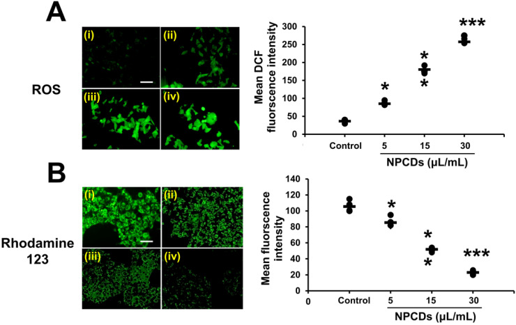
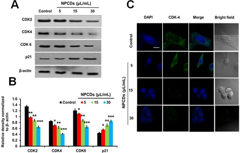
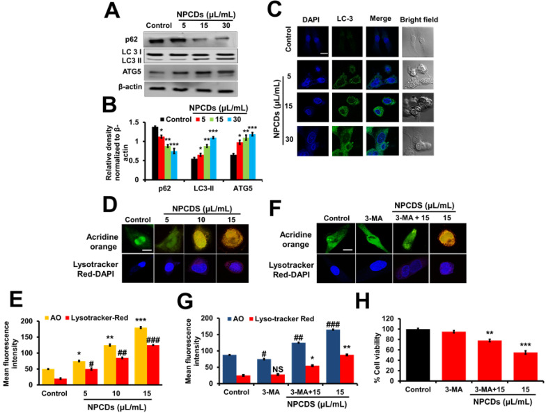
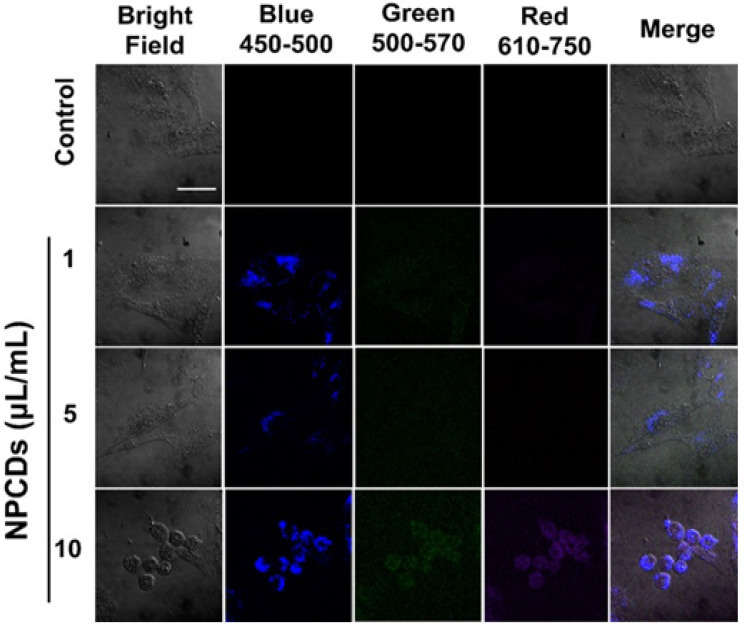
The present study reports the multifunctional anticancer activity against B16F10 melanoma cancer cells and the bioimaging ability of fluorescent nitrogen-phosphorous-doped carbon dots (NPCDs). The NPCDs were synthesized using a single-step, thermal treatment and were characterized by TEM, XPS, fluorescence and UV-Vis spectroscopy, and FTIR analysis. The anticancer efficacy of NPCDs was confirmed by using cell viability assay, morphological evaluation, fluorescent live-dead cell assay, mitochondrial potential assay, ROS production, RT-PCR, western-blot analysis, siRNA transfection, and cellular bioimaging ability. The NPCDs inhibited the proliferation of B16F10 melanoma cancer cells after 24 h of treatment and induced apoptosis, as confirmed by the presence of fragmented nuclei, reduced mitochondrial membrane potential, and elevated levels of reactive oxygen species. The NPCDs treatment further elevated the levels of pro-apoptotic factors and down-regulated the level of Bcl2 (B-cell lymphoma 2) that weakened the mitochondrial membrane, and activated proteases such as caspases. Treatment with NPCDs also resulted in dose-dependent cell cycle arrest, as indicated by reduced cyclin-dependent kinase (CDK)-2, -4, and -6 protein levels and an enhanced level of p21. More importantly, the NPCDs induced the activation of autophagy by upregulating the protein expression levels of LC3-II and ATG-5 (autophagy-related-5) and by downregulating p62 level, validated by knockdown of ATG-5. Additionally, owing to their excellent luminescence property, these NPCDs were also applicable in cellular bioimaging, as evidenced by the microscopic fluorescence imaging of B16F10 melanoma cells. Based on these findings, we conclude that our newly synthesized NPCDs induced cell cycle arrest, autophagy, and apoptosis in B16F10 melanoma cells and presented good cellular bioimaging capability.
本研究报告了荧光氮磷掺杂碳点(NPCDs)对 B16F10 黑色素瘤癌细胞的多功能抗癌活性和生物成像能力。NPCDs 是通过一步法、热处理合成的,并通过 TEM、XPS、荧光和紫外-可见光谱以及 FTIR 分析进行了表征。通过细胞活力测定、形态评估、荧光死活细胞测定、线粒体电位测定、ROS 产生、RT-PCR、western blot 分析、siRNA 转染和细胞生物成像能力证实了 NPCDs 的抗癌功效。NPCDs 在处理 24 小时后抑制了 B16F10 黑色素瘤癌细胞的增殖,并诱导了细胞凋亡,这表现在核碎片的出现、线粒体膜电位的降低以及活性氧水平的升高。NPCDs 处理还进一步提高了促凋亡因子的水平,并下调了 Bcl2(B 细胞淋巴瘤 2)的水平,从而削弱了线粒体膜,并激活了蛋白酶如半胱天冬酶。NPCDs 的处理还导致了细胞周期依赖性的细胞周期停滞,这表现为细胞周期蛋白依赖性激酶(CDK)-2、-4 和 -6 蛋白水平降低,p21 水平升高。更重要的是,NPCDs 通过上调 LC3-II 和 ATG-5(自噬相关蛋白-5)的蛋白表达水平和下调 p62 水平诱导自噬的激活,这通过 ATG-5 的敲低得到验证。此外,由于其优异的发光性能,这些 NPCDs 也适用于细胞生物成像,这从 B16F10 黑色素瘤细胞的微观荧光成像中得到了证明。基于这些发现,我们得出结论,我们新合成的 NPCDs 诱导了 B16F10 黑色素瘤细胞的细胞周期停滞、自噬和凋亡,并具有良好的细胞生物成像能力。