• 文献检索
  • 文档翻译
  • 深度研究
  • 学术资讯
  • Suppr Zotero 插件Zotero 插件
  • 邀请有礼
  • 套餐&价格
  • 历史记录
应用&插件
Suppr Zotero 插件Zotero 插件浏览器插件Mac 客户端Windows 客户端微信小程序
定价
高级版会员购买积分包购买API积分包
服务
文献检索文档翻译深度研究API 文档MCP 服务
关于我们
关于 Suppr公司介绍联系我们用户协议隐私条款
关注我们

Suppr 超能文献

核心技术专利:CN118964589B侵权必究
粤ICP备2023148730 号-1Suppr @ 2026

文献检索

告别复杂PubMed语法,用中文像聊天一样搜索,搜遍4000万医学文献。AI智能推荐,让科研检索更轻松。

立即免费搜索

文件翻译

保留排版,准确专业,支持PDF/Word/PPT等文件格式,支持 12+语言互译。

免费翻译文档

深度研究

AI帮你快速写综述,25分钟生成高质量综述,智能提取关键信息,辅助科研写作。

立即免费体验

中国浙江省温州市出院 COVID-19 患者的胸部计算机断层扫描和临床随访。

Chest Computed Tomography and Clinical Follow-Up of Discharged Patients with COVID-19 in Wenzhou City, Zhejiang, China.

机构信息

Department of Radiation Oncology, National Cancer Center/National Clinical Research Center for Cancer/Cancer Hospital and Shenzhen Hospital, Chinese Academy of Medical Sciences and Peking Union Medical College, Shenzhen, China.

Department of Respiration, Zhongshan Hospital, FuDan University, Shanghai, China.

出版信息

Ann Am Thorac Soc. 2020 Oct;17(10):1231-1237. doi: 10.1513/AnnalsATS.202004-324OC.

DOI:10.1513/AnnalsATS.202004-324OC
PMID:32692945
原文链接:https://pmc.ncbi.nlm.nih.gov/articles/PMC7640627/
Abstract

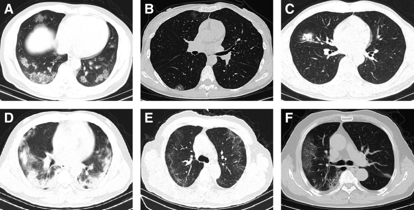
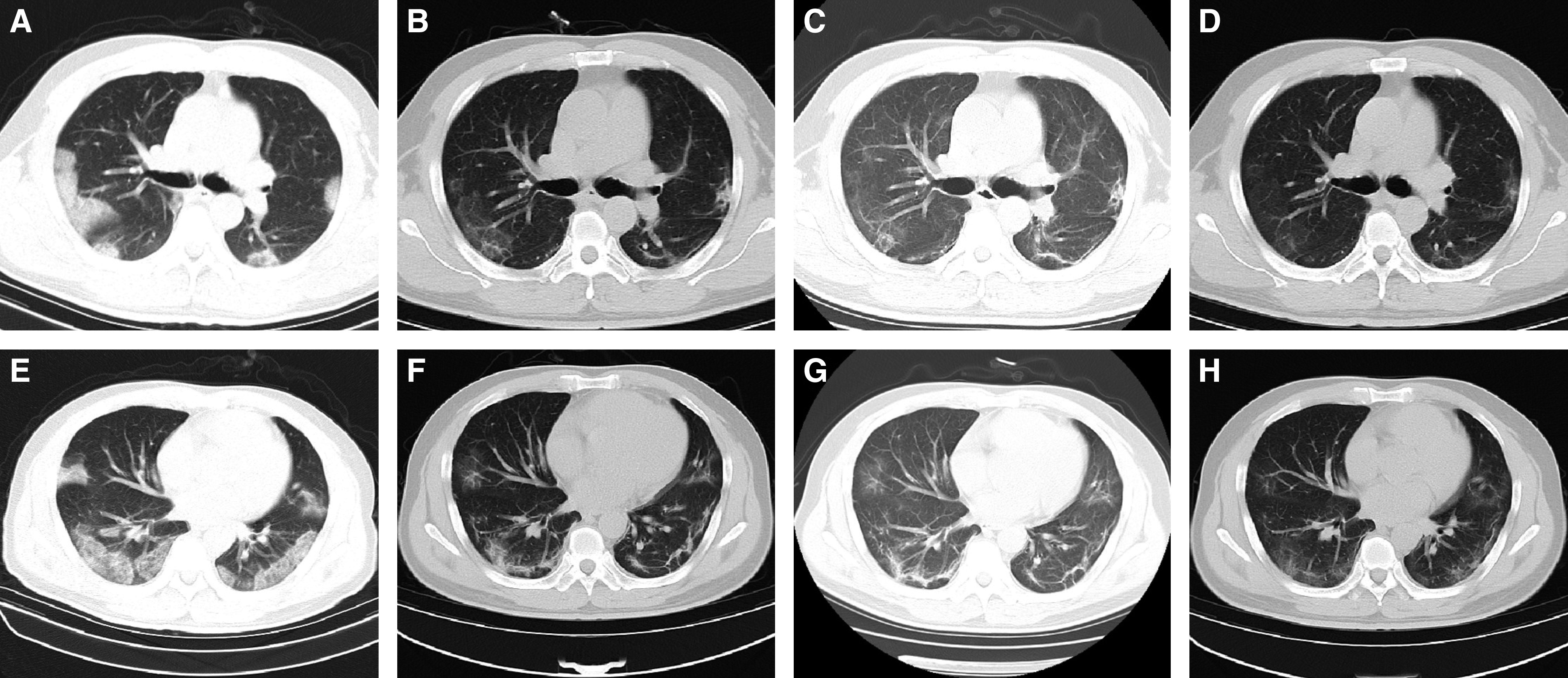
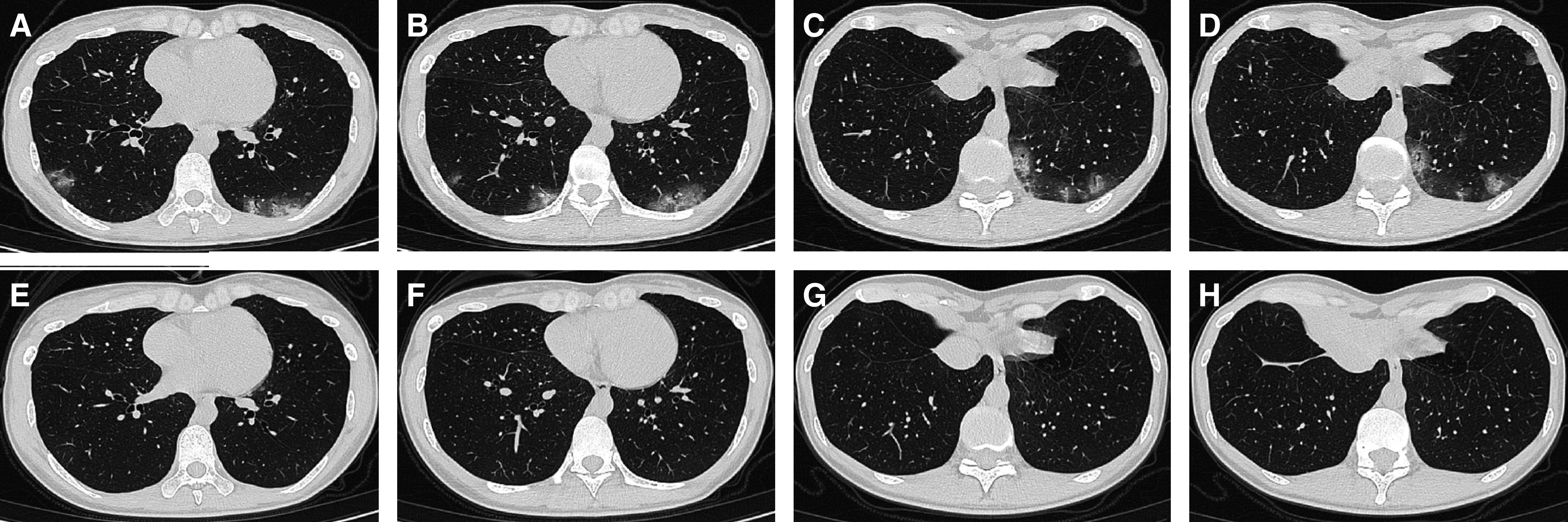
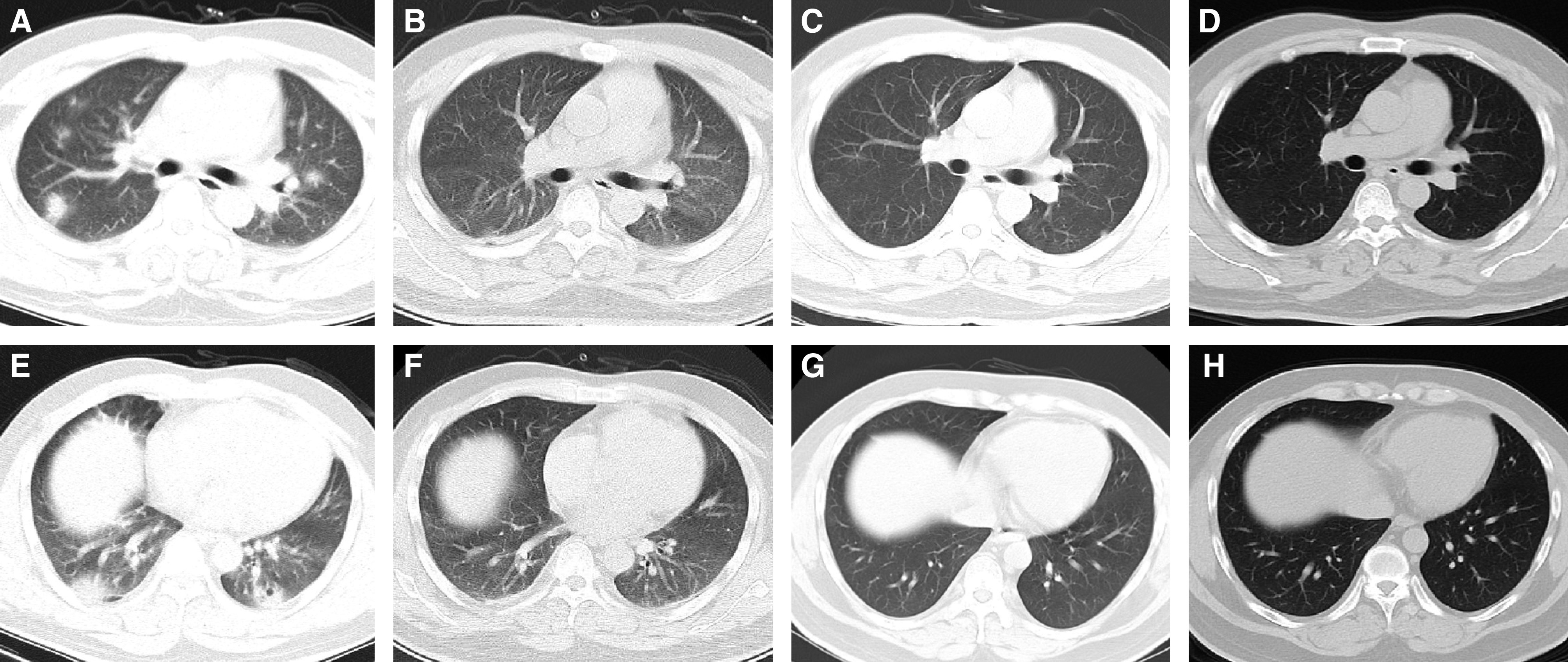
Many clinical studies have focused on the epidemiological and clinical characteristics of inpatients with coronavirus disease (COVID-19). However, there are few reports about the clinical follow-up of discharged patients. To describe the follow-up of patients with COVID-19 in Wenzhou City, Zhejiang, China. We retrospectively reviewed 4-week follow-ups in patients with COVID-19, including computed tomographic (CT) chest scanning, blood testing, and oropharyngeal-swab testing for severe acute respiratory syndrome coronavirus 2 (SARS-CoV-2) ribonucleic acid. The chest CT scans and blood tests were performed on the last day before discharge and 2 weeks and 4 weeks after discharge. The oropharyngeal-swab tests were performed at both 1 week and 2 weeks after discharge. Fifty-one patients with common COVID-19 were enrolled in the study. All the CT and clinical data were collected between January 23 and March 28, 2020. Compared with the abnormalities found on the the last CT scans before discharge, the abnormalities in the lungs at the first and second follow-ups after discharge had been gradually absorbed. The cases with focal ground-glass opacity were reduced from 17.7% to 9.8% of cases. The cases with multiple ground-glass opacities decreased from 80.4% to 23.5%. The cases with consolidation were reduced from 49.0% to 2.0%. The cases with interlobular septal thickening were reduced from 80.4% to 35.3%. The cases with subpleural lines were reduced from 29.4% to 7.8%. The cases with irregular lines were reduced from 41.2% to 15.7%. The lung lesions of 25.5% patients were shown to be fully absorbed on the first CT scans after discharge, and the rate of lung recovery increased to 64.7% after the second follow-up. Nucleic-acid test results became recurrently positive in 17.6% of discharged patients, of whom only 33.3% complained of clinical symptoms. There were no differences in the characteristics of the last CT scans before discharge between the patients with recurrently positive test results and the patients with negative test results. The lung damage was fully absorbed in 55.6% of discharged patients with recurrence of positive test results for SARS-CoV-2 ribonucleic acid. The lung damage due to COVID-19 could be reversible for patients with common COVID-19. A few cases showed recurring positive results of nucleic-acid tests after discharge.

摘要

许多临床研究都集中在冠状病毒病(COVID-19)住院患者的流行病学和临床特征上。然而,关于出院患者的临床随访报告很少。本研究旨在描述中国浙江省温州市 COVID-19 患者的随访情况。我们回顾性分析了 4 周的 COVID-19 出院患者随访,包括胸部计算机断层扫描(CT)、血液检测和咽拭子检测严重急性呼吸综合征冠状病毒 2(SARS-CoV-2)的核糖核酸。胸部 CT 扫描和血液检查分别在出院前最后一天、出院后 2 周和 4 周进行。咽拭子检查分别在出院后第 1 周和第 2 周进行。本研究纳入了 51 例普通 COVID-19 患者。所有 CT 和临床数据均于 2020 年 1 月 23 日至 3 月 28 日收集。与出院前最后一次 CT 扫描的异常相比,出院后第一次和第二次随访时肺部的异常逐渐吸收。局灶性磨玻璃影的病例从 17.7%减少到 9.8%。多发性磨玻璃影的病例从 80.4%减少到 23.5%。实变的病例从 49.0%减少到 2.0%。间隔增厚的病例从 80.4%减少到 35.3%。胸膜下线的病例从 29.4%减少到 7.8%。不规则线的病例从 41.2%减少到 15.7%。25.5%的患者出院后首次 CT 扫描显示肺部病变完全吸收,第二次随访后肺部恢复率增加至 64.7%。出院患者中有 17.6%的核酸检测结果呈复阳,其中仅 33.3%有临床症状。复阳患者与阴性患者出院前最后一次 CT 扫描的特征无差异。55.6%的复阳患者 SARS-CoV-2 核糖核酸检测结果完全吸收。普通 COVID-19 患者的肺部损伤可能是可逆的。少数患者出院后 SARS-CoV-2 核糖核酸检测结果出现反复阳性。

https://cdn.ncbi.nlm.nih.gov/pmc/blobs/12b6/7640627/0cd15b9ab6f6/AnnalsATS.202004-324OCf4.jpg
https://cdn.ncbi.nlm.nih.gov/pmc/blobs/12b6/7640627/88e156758255/AnnalsATS.202004-324OCf1.jpg
https://cdn.ncbi.nlm.nih.gov/pmc/blobs/12b6/7640627/bf7c75065df2/AnnalsATS.202004-324OCf2.jpg
https://cdn.ncbi.nlm.nih.gov/pmc/blobs/12b6/7640627/6dfebd857560/AnnalsATS.202004-324OCf3.jpg
https://cdn.ncbi.nlm.nih.gov/pmc/blobs/12b6/7640627/0cd15b9ab6f6/AnnalsATS.202004-324OCf4.jpg

相似文献

1
Chest Computed Tomography and Clinical Follow-Up of Discharged Patients with COVID-19 in Wenzhou City, Zhejiang, China.中国浙江省温州市出院 COVID-19 患者的胸部计算机断层扫描和临床随访。
Ann Am Thorac Soc. 2020 Oct;17(10):1231-1237. doi: 10.1513/AnnalsATS.202004-324OC.
2
An uncommon manifestation of COVID-19 pneumonia on CT scan with small cavities in the lungs: A case report.新冠肺炎肺部CT扫描显示肺部有小空洞的罕见表现:一例报告。
Medicine (Baltimore). 2020 Jul 10;99(28):e21240. doi: 10.1097/MD.0000000000021240.
3
Imaging and clinical features of patients with 2019 novel coronavirus SARS-CoV-2.新型冠状病毒 2019 年 SARS-CoV-2 患者的影像学和临床特征。
Eur J Nucl Med Mol Imaging. 2020 May;47(5):1275-1280. doi: 10.1007/s00259-020-04735-9. Epub 2020 Feb 28.
4
Radiographic and Clinical Features of Children With Coronavirus Disease (COVID-19) Pneumonia.儿童新型冠状病毒肺炎(COVID-19)肺炎的影像学和临床特征。
Indian Pediatr. 2020 May 15;57(5):423-426. doi: 10.1007/s13312-020-1816-8. Epub 2020 Apr 7.
5
[Clinical analysis of 150 cases of 2019 novel coronavirus infection in Nanyang City, Henan Province].[河南省南阳市150例2019新型冠状病毒感染病例的临床分析]
Zhonghua Jie He He Hu Xi Za Zhi. 2020 Jun 12;43(6):503-508. doi: 10.3760/cma.j.cn112147-20200224-00168.
6
Clinical and computed tomographic imaging features of novel coronavirus pneumonia caused by SARS-CoV-2.SARS-CoV-2 引起的新型冠状病毒肺炎的临床和计算机断层扫描影像学特征。
J Infect. 2020 Apr;80(4):394-400. doi: 10.1016/j.jinf.2020.02.017. Epub 2020 Feb 25.
7
Temporal Changes of CT Findings in 90 Patients with COVID-19 Pneumonia: A Longitudinal Study.90 例 COVID-19 肺炎患者 CT 表现的时间变化:一项纵向研究。
Radiology. 2020 Aug;296(2):E55-E64. doi: 10.1148/radiol.2020200843. Epub 2020 Mar 19.
8
Clinical features and outcomes of discharged coronavirus disease 2019 patients: a prospective cohort study.出院的 2019 年冠状病毒病患者的临床特征和结局:一项前瞻性队列研究。
QJM. 2020 Sep 1;113(9):657-665. doi: 10.1093/qjmed/hcaa178.
9
Clinical characteristics and imaging manifestations of the 2019 novel coronavirus disease (COVID-19):A multi-center study in Wenzhou city, Zhejiang, China.中国浙江省温州市多中心研究:2019 年新型冠状病毒病(COVID-19)的临床特征和影像学表现。
J Infect. 2020 Apr;80(4):388-393. doi: 10.1016/j.jinf.2020.02.016. Epub 2020 Feb 26.
10
Preliminary CT findings of coronavirus disease 2019 (COVID-19).新型冠状病毒肺炎(COVID-19)的初步 CT 影像学表现。
Clin Imaging. 2020 Sep;65:124-132. doi: 10.1016/j.clinimag.2020.04.042. Epub 2020 May 12.

引用本文的文献

1
Comparison of Tomographic Findings in Patients with Influenza A (H1N1) and COVID-19.甲型H1N1流感与新型冠状病毒肺炎患者的断层扫描结果比较
Diagnostics (Basel). 2025 Jun 4;15(11):1430. doi: 10.3390/diagnostics15111430.
2
Early pulmonary fibrosis-like changes between delta and pre-delta periods in patients with severe COVID-19 pneumonia on mechanical ventilation.机械通气治疗的重症 COVID-19 肺炎患者在 delta 期和 pre-delta 期之间出现早期肺纤维化样改变。
Sci Rep. 2024 Oct 30;14(1):26101. doi: 10.1038/s41598-024-77405-7.
3
Clinico-radiological and pulmonary function assessment of post-COVID-19 patients with respiratory symptoms.

本文引用的文献

1
Limits of Detection of 6 Approved RT-PCR Kits for the Novel SARS-Coronavirus-2 (SARS-CoV-2).6种获批的新型严重急性呼吸综合征冠状病毒2(SARS-CoV-2)逆转录聚合酶链反应(RT-PCR)试剂盒的检测限
Clin Chem. 2020 Jul 1;66(7):977-979. doi: 10.1093/clinchem/hvaa099.
2
Virological assessment of hospitalized patients with COVID-2019.住院 COVID-19 患者的病毒学评估。
Nature. 2020 May;581(7809):465-469. doi: 10.1038/s41586-020-2196-x. Epub 2020 Apr 1.
3
SARS-CoV-2 turned positive in a discharged patient with COVID-19 arouses concern regarding the present standards for discharge.
有呼吸道症状的新冠后患者的临床影像学和肺功能评估
J Family Med Prim Care. 2024 Aug;13(8):2912-2920. doi: 10.4103/jfmpc.jfmpc_1721_23. Epub 2024 Jul 26.
4
Characteristics and determinants of pulmonary long COVID.肺长新冠的特征和决定因素。
JCI Insight. 2024 Apr 23;9(10):e177518. doi: 10.1172/jci.insight.177518.
5
Research progress of post-acute sequelae after SARS-CoV-2 infection.新型冠状病毒感染后急性后遗症的研究进展。
Cell Death Dis. 2024 Apr 11;15(4):257. doi: 10.1038/s41419-024-06642-5.
6
Optical Coherence Tomography Angiography Assessment of the Optic Nerve Head in Patients Hospitalized Due to COVID-19 Bilateral Pneumonia.因 COVID-19 双侧肺炎住院患者视神经头的光学相干断层扫描血管造影评估。
Medicina (Kaunas). 2024 Mar 19;60(3):502. doi: 10.3390/medicina60030502.
7
Persistence of long COVID symptoms in COVID-19 survivors worldwide and its potential pathogenesis - A systematic review and meta-analysis.全球新冠病毒感染者康复后长期新冠症状的持续情况及其潜在发病机制——一项系统综述和荟萃分析
Narra J. 2021 Aug;1(2):e36. doi: 10.52225/narraj.v1i2.36. Epub 2021 Aug 1.
8
Fatigue has a prominent impact on health lasting 12-weeks after COVID-19 infection.感染 COVID-19 12 周后,疲劳对健康的影响显著。
Malawi Med J. 2023 Jun;35(2):124-129. doi: 10.4314/mmj.v35i2.9.
9
Clinical Characterization of the Persistent COVID-19 Symptoms: A Descriptive Observational Study in Primary Care.持续性 COVID-19 症状的临床特征:初级保健中的描述性观察研究。
J Prim Care Community Health. 2023 Jan-Dec;14:21501319231208283. doi: 10.1177/21501319231208283.
10
Clinical and pulmonary function analysis in long-COVID revealed that long-term pulmonary dysfunction is associated with vascular inflammation pathways and metabolic syndrome.新冠后长期症状的临床和肺功能分析显示,长期肺功能障碍与血管炎症途径和代谢综合征有关。
Front Med (Lausanne). 2023 Oct 6;10:1271863. doi: 10.3389/fmed.2023.1271863. eCollection 2023.
出院的 COVID-19 患者新冠病毒检测呈阳性,这引发了人们对目前出院标准的关注。
Int J Infect Dis. 2020 Aug;97:212-214. doi: 10.1016/j.ijid.2020.03.007. Epub 2020 Mar 19.
4
Post-discharge surveillance and positive virus detection in two medical staff recovered from coronavirus disease 2019 (COVID-19), China, January to February 2020.2020 年 1 月至 2 月,中国两例新冠肺炎(COVID-19)治愈医务人员出院后随访和病毒阳性检出情况。
Euro Surveill. 2020 Mar;25(10). doi: 10.2807/1560-7917.ES.2020.25.10.2000191.
5
Recurrence of positive SARS-CoV-2 RNA in COVID-19: A case report.COVID-19 中 SARS-CoV-2 RNA 复阳:一例报告。
Int J Infect Dis. 2020 Apr;93:297-299. doi: 10.1016/j.ijid.2020.03.003. Epub 2020 Mar 5.
6
A pneumonia outbreak associated with a new coronavirus of probable bat origin.一种新型冠状病毒引发的肺炎疫情,该病毒可能来源于蝙蝠。
Nature. 2020 Mar;579(7798):270-273. doi: 10.1038/s41586-020-2012-7. Epub 2020 Feb 3.
7
Nowcasting and forecasting the potential domestic and international spread of the 2019-nCoV outbreak originating in Wuhan, China: a modelling study.实时预测和预报源自中国武汉的 2019-nCoV 疫情在国内和国际的潜在传播:一项建模研究。
Lancet. 2020 Feb 29;395(10225):689-697. doi: 10.1016/S0140-6736(20)30260-9. Epub 2020 Jan 31.
8
Genomic characterisation and epidemiology of 2019 novel coronavirus: implications for virus origins and receptor binding.新冠病毒的基因组特征和流行病学:对病毒起源和受体结合的影响。
Lancet. 2020 Feb 22;395(10224):565-574. doi: 10.1016/S0140-6736(20)30251-8. Epub 2020 Jan 30.
9
A Novel Coronavirus from Patients with Pneumonia in China, 2019.2019 年中国肺炎患者中的一种新型冠状病毒。
N Engl J Med. 2020 Feb 20;382(8):727-733. doi: 10.1056/NEJMoa2001017. Epub 2020 Jan 24.
10
The continuing 2019-nCoV epidemic threat of novel coronaviruses to global health - The latest 2019 novel coronavirus outbreak in Wuhan, China.新型冠状病毒持续的2019 - nCoV疫情对全球健康构成威胁——中国武汉最新的2019新型冠状病毒爆发。
Int J Infect Dis. 2020 Feb;91:264-266. doi: 10.1016/j.ijid.2020.01.009. Epub 2020 Jan 14.