Rytelewski Mateusz, Harutyunyan Karine, Baran Natalia, Mallampati Saradhi, Zal M Anna, Cavazos Antonio, Butler Jason M, Konoplev Sergej, El Khatib Mirna, Plunkett Shane, Marszalek Joseph R, Andreeff Michael, Zal Tomasz, Konopleva Marina
Department of Immunology, The University of Texas MD Anderson Cancer Center, Houston, TX, United States.
Department of Leukemia, The University of Texas MD Anderson Cancer Center, Houston, TX, United States.
Front Oncol. 2020 Jun 30;10:991. doi: 10.3389/fonc.2020.00991. eCollection 2020.
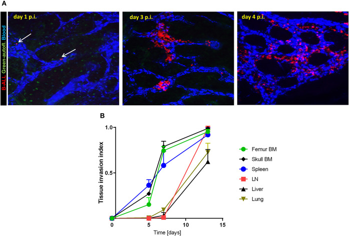
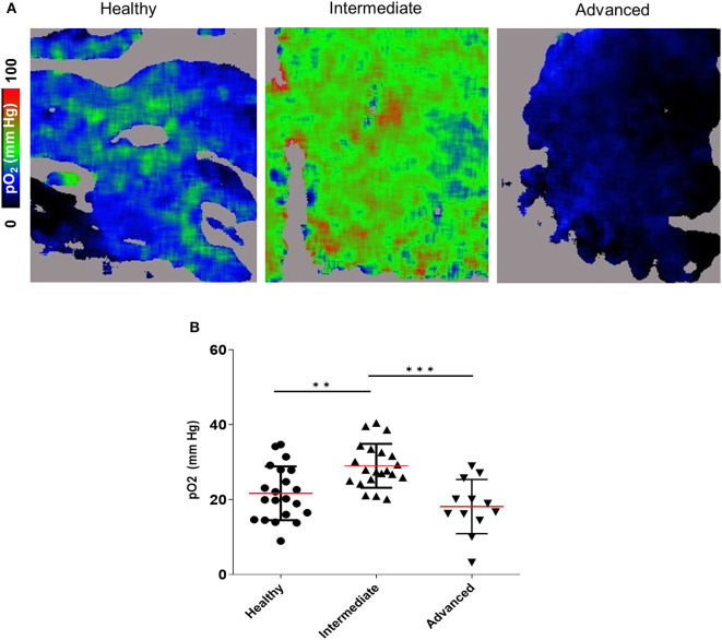
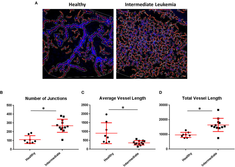
Abnormally low level of interstitial oxygen, or hypoxia, is a hallmark of tumor microenvironment and a known promoter of cancer chemoresistance. Inside a solid tumor mass, the hypoxia stems largely from inadequate supply of oxygenated blood through sparse or misshapen tumor vasculature whilst oxygen utilization rates are low in typical tumor's glycolytic metabolism. In acute leukemias, however, markers of intracellular hypoxia such as increased pimonidazole adduct staining and HIF-1α stabilization are observed in advanced leukemic bone marrows (BM) despite an increase in BM vasculogenesis. We utilized intravital fast scanning two-photon phosphorescence lifetime imaging microscopy (FaST-PLIM) in a BCR-ABL B-ALL mouse model to image the extracellular oxygen concentrations (pO) in leukemic BM, and we related the extracellular oxygen levels to intracellular hypoxia, vascular markers and local leukemia burden. We observed a transient increase in BM pO in initial disease stages with intermediate leukemia BM burden, which correlated with an expansion of blood-carrying vascular network in the BM. Yet, we also observed increased formation of intracellular pimonidazole adducts in leukemic BM at the same time. This intermediate stage was followed by a significant decrease of extracellular pO and further increase of intracellular hypoxia as leukemia cellularity overwhelmed BM in disease end-stage. Remarkably, treatment of leukemic mice with IACS-010759, a pharmacological inhibitor of mitochondrial Complex I, substantially increased pO in the BM with advanced B-ALL, and it alleviated intracellular hypoxia reported by pimonidazole staining. High rates of oxygen consumption by B-ALL cells were confirmed by Seahorse assay including in cells. Our results suggest that B-ALL expansion in BM is associated with intense oxidative phosphorylation (OxPhos) leading to the onset of metabolic BM hypoxia despite increased BM vascularization. Targeting mitochondrial respiration may be a novel approach to counteract BM hypoxia in B-ALL and, possibly, tumor hypoxia in other OxPhos-reliant malignancies.
间质氧水平异常低下,即缺氧,是肿瘤微环境的一个标志,也是已知的癌症化疗耐药性促进因素。在实体肿瘤块内部,缺氧主要源于通过稀疏或畸形的肿瘤血管系统供应的含氧血液不足,而在典型的肿瘤糖酵解代谢中,氧利用率较低。然而,在急性白血病中,尽管骨髓血管生成增加,但在晚期白血病骨髓(BM)中仍观察到细胞内缺氧标志物,如匹莫硝唑加合物染色增加和HIF-1α稳定。我们在BCR-ABL B-ALL小鼠模型中利用活体快速扫描双光子磷光寿命成像显微镜(FaST-PLIM)对白血病骨髓中的细胞外氧浓度(pO)进行成像,并将细胞外氧水平与细胞内缺氧、血管标志物和局部白血病负担相关联。我们观察到在白血病骨髓负担中等的疾病初始阶段,骨髓pO出现短暂升高,这与骨髓中携带血液的血管网络扩张相关。然而,我们同时也观察到白血病骨髓中细胞内匹莫硝唑加合物的形成增加。在疾病终末期,随着白血病细胞数量超过骨髓,这个中间阶段之后是细胞外pO显著下降和细胞内缺氧进一步增加。值得注意的是,用线粒体复合物I的药理学抑制剂IACS-010759治疗白血病小鼠,可显著提高晚期B-ALL骨髓中的pO,并减轻匹莫硝唑染色报告的细胞内缺氧。通过海马实验证实了B-ALL细胞的高耗氧率,包括在细胞中。我们的结果表明,尽管骨髓血管化增加,但骨髓中B-ALL的扩张与强烈的氧化磷酸化(OxPhos)相关,导致代谢性骨髓缺氧的发生。靶向线粒体呼吸可能是对抗B-ALL骨髓缺氧以及其他依赖OxPhos的恶性肿瘤中肿瘤缺氧的一种新方法。