Laboratory for Transplantation and Regenerative Medicine, Sahlgrenska Academy, University of Gothenburg, Kvinnokliniken, Blå stråket 6, SE-413 45, Göteborg, Sweden.
Department of Obstetrics and Gynecology, Sahlgrenska Academy, University of Gothenburg, Gothenburg, Sweden.
Reprod Biol Endocrinol. 2020 Jul 23;18(1):75. doi: 10.1186/s12958-020-00630-y.
Fertility preservation is particularly challenging in young women diagnosed with hematopoietic cancers, as transplantation of cryopreserved ovarian cortex in these women carries the risk for re-introducing cancer cells. Therefore, the construction of a bioengineered ovary that can accommodate isolated small follicles was proposed as an alternative to minimize the risk of malignancy transmission. Various options for viable bioengineered scaffolds have been reported in the literature. Previously, we reported three protocols for producing mouse ovarian scaffolds with the decellularization technique. The present study examined these scaffolds further, specifically with regards to their extracellular composition, biocompatibility and ability to support recellularization with mesenchymal stem cells.
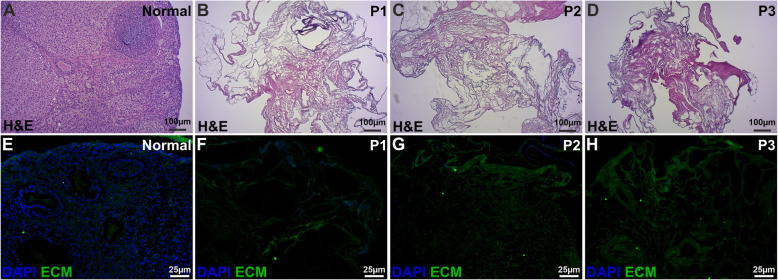
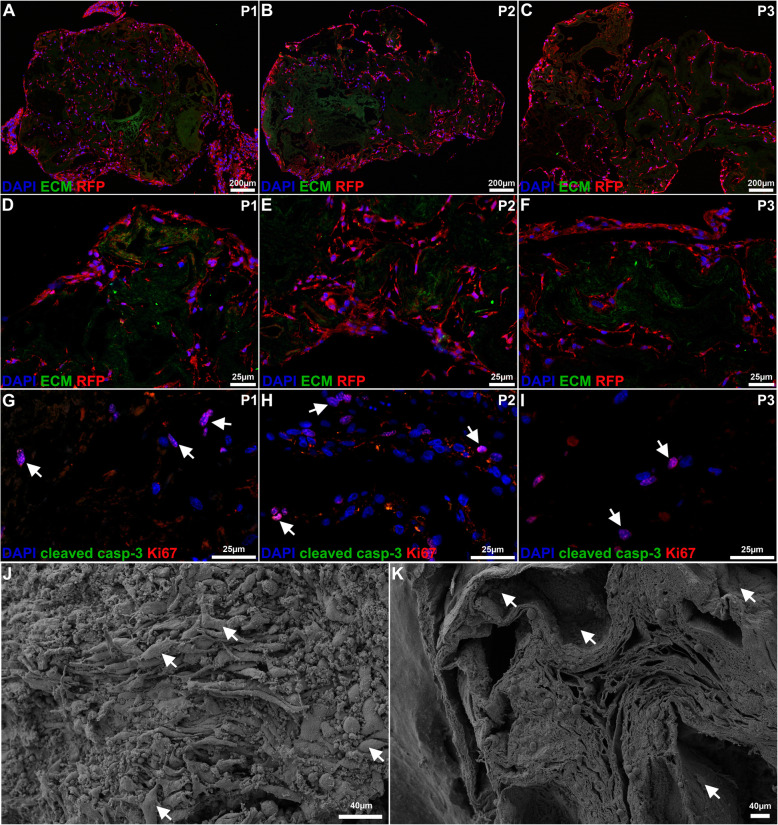
Three decellularization protocols based on 0.5% sodium dodecyl sulfate (Protocol 1; P1), or 2% sodium deoxycholate (P2), or a combination of the two detergents (P3) were applied to produce three types of scaffolds. The levels of collagen, elastin and sulfated glycosaminoglycans (sGAGs) were quantified in the remaining extracellular matrix. Detailed immunofluorescence and scanning electron microscopy imaging were conducted to assess the morphology and recellularization efficiency of the constructs after 14 days in vitro utilizing red fluorescent protein-labelled mesenchymal stem cells.
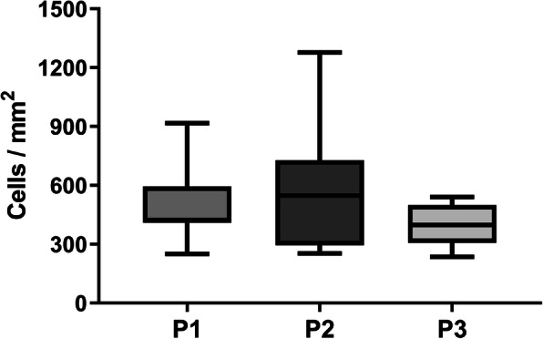
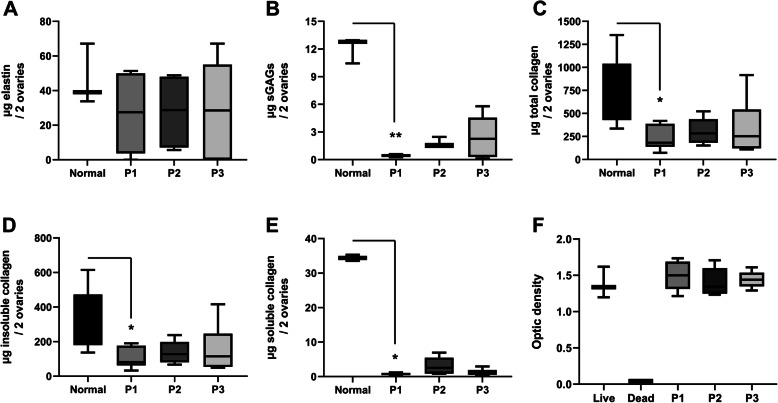
All protocols efficiently removed the DNA while the elastin content was not significantly reduced during the procedures. The SDS-protocol (P1) reduced the sGAG and the collagen content more than the SDC-protocol (P2). All scaffolds were biocompatible and recellularization was successful, particularly in several P2-derived scaffolds. The cells were extensively distributed throughout the constructs, with a denser distribution observed towards the ovarian cortex. The cell density was not significantly different (400 to 550 cells/mm) between scaffold types. However, there was a tendency towards a higher cell density in the SDC-derived constructs. Scanning electron microscope images showed fibrous scaffolds with a dense repopulated surface structure.
While there were differences in the key structural macromolecules between protocols, all scaffolds were biocompatible and showed effective recellularization. The results indicate that our SDC-protocol might be better than our SDS-protocol. However, additional studies are necessary to determine their suitability for attachment of small follicles and folliculogenesis.
对于被诊断患有血液系统癌症的年轻女性,生育力保存极具挑战性,因为移植冷冻卵巢皮质会有重新引入癌细胞的风险。因此,构建一个可容纳分离的小卵泡的生物工程卵巢被提议作为一种替代方案,以最大限度地降低恶性传播的风险。文献中已经报道了各种可行的生物工程支架的选择。此前,我们报道了三种使用脱细胞技术生产小鼠卵巢支架的方案。本研究进一步研究了这些支架,特别是它们的细胞外成分、生物相容性以及支持间充质干细胞再细胞化的能力。
基于 0.5%十二烷基硫酸钠(方案 1;P1)或 2%脱氧胆酸钠(P2)或两种去污剂的组合(P3)应用了三种脱细胞方案来生产三种类型的支架。在剩余的细胞外基质中定量了胶原蛋白、弹性蛋白和硫酸化糖胺聚糖(sGAG)的水平。详细的免疫荧光和扫描电子显微镜成像用于评估在体外培养 14 天后构建体的形态和再细胞化效率,利用红色荧光蛋白标记的间充质干细胞。
所有方案均能有效去除 DNA,而弹性蛋白含量在处理过程中没有明显减少。SDS 方案(P1)比 SDC 方案(P2)更能减少 sGAG 和胶原蛋白含量。所有支架均具有生物相容性,再细胞化成功,特别是在几个 P2 衍生的支架中。细胞广泛分布在整个构建体中,靠近卵巢皮质的分布更为密集。细胞密度在支架类型之间没有显著差异(400 到 550 个细胞/mm)。然而,SDC 衍生的构建体中细胞密度有增加的趋势。扫描电子显微镜图像显示出具有密集再殖表面结构的纤维状支架。
尽管方案之间在关键结构大分子方面存在差异,但所有支架均具有生物相容性,并表现出有效的再细胞化。结果表明,我们的 SDC 方案可能优于我们的 SDS 方案。然而,需要进一步的研究来确定它们是否适合附着小卵泡和卵泡发生。