Department of Biomedical Engineering, College of Engineering, Peking University, Beijing, 100871, China.
School of Applied and Engineering Physics, Cornell University, Ithaca, 14853, USA.
Nat Commun. 2020 Jul 24;11(1):3699. doi: 10.1038/s41467-020-17546-1.
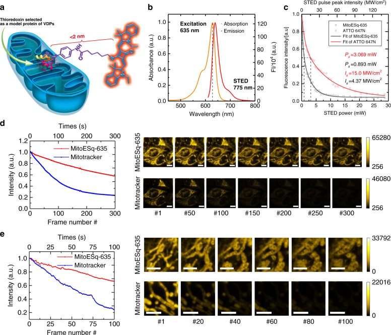
Mitochondria play a critical role in generating energy to support the entire lifecycle of biological cells, yet it is still unclear how their morphological structures evolve to regulate their functionality. Conventional fluorescence microscopy can only provide ~300 nm resolution, which is insufficient to visualize mitochondrial cristae. Here, we developed an enhanced squaraine variant dye (MitoESq-635) to study the dynamic structures of mitochondrial cristae in live cells with a superresolution technique. The low saturation intensity and high photostability of MitoESq-635 make it ideal for long-term, high-resolution (stimulated emission depletion) STED nanoscopy. We performed time-lapse imaging of the mitochondrial inner membrane over 50 min (3.9 s per frame, with 71.5 s dark recovery) in living HeLa cells with a resolution of 35.2 nm. The forms of the cristae during mitochondrial fusion and fission can be clearly observed. Our study demonstrates the emerging capability of optical STED nanoscopy to investigate intracellular physiological processes with nanoscale resolution for an extended period of time.
线粒体在为生物细胞的整个生命周期提供能量方面发挥着关键作用,但线粒体形态结构如何进化以调节其功能仍不清楚。传统的荧光显微镜只能提供约 300nm 的分辨率,不足以可视化线粒体嵴。在这里,我们开发了一种增强的方酸变体染料(MitoESq-635),并用超分辨率技术研究活细胞中线粒体嵴的动态结构。MitoESq-635 的低饱和强度和高光稳定性使其成为用于长期、高分辨率(受激发射损耗)STED 纳米显微镜的理想选择。我们在活 HeLa 细胞中以 35.2nm 的分辨率进行了线粒体内膜的延时成像(每帧 3.9s,暗恢复 71.5s),时间长达 50min(3.9s 每帧,71.5s 暗恢复)。可以清楚地观察到线粒体融合和裂变过程中嵴的形态。我们的研究表明,光学 STED 纳米显微镜具有在纳米尺度上长时间研究细胞内生理过程的新兴能力。