Bottanelli Francesca, Kromann Emil B, Allgeyer Edward S, Erdmann Roman S, Wood Baguley Stephanie, Sirinakis George, Schepartz Alanna, Baddeley David, Toomre Derek K, Rothman James E, Bewersdorf Joerg
Department of Cell Biology, Yale University School of Medicine, New Haven, Connecticut 06520, USA.
Department of Biomedical Engineering, Yale University, New Haven, Connecticut 06520, USA.
Nat Commun. 2016 Mar 4;7:10778. doi: 10.1038/ncomms10778.
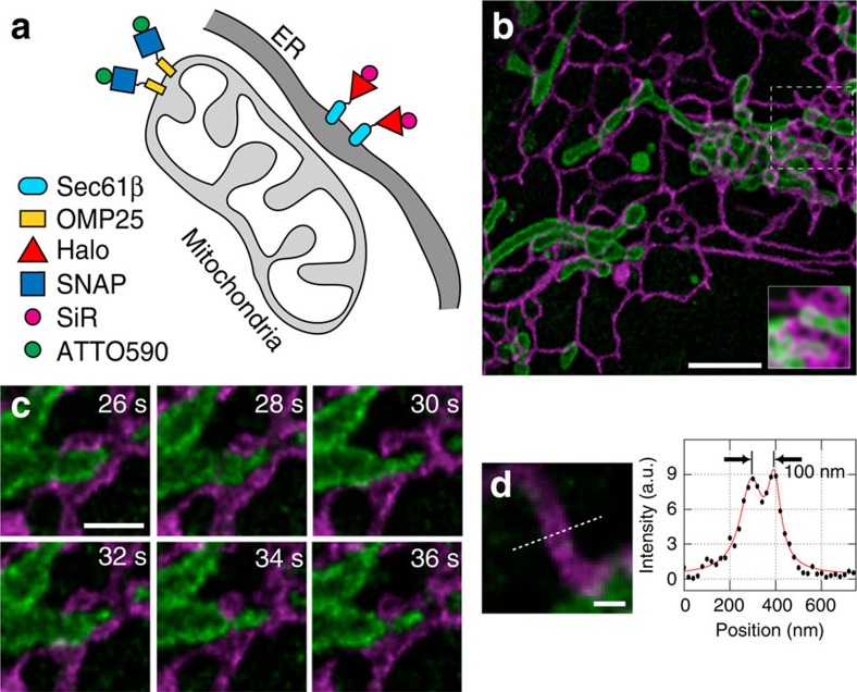
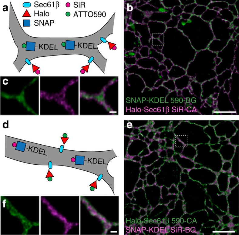
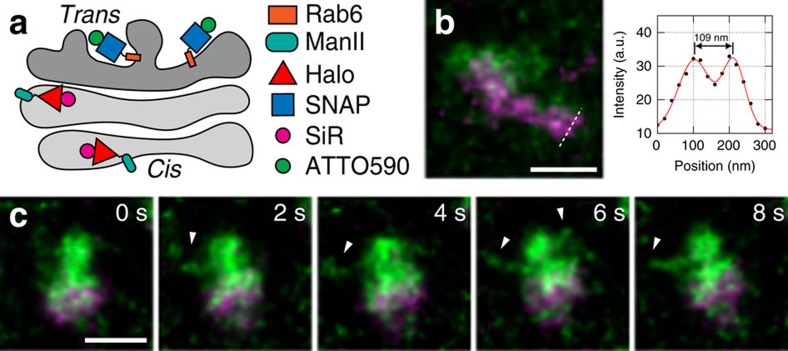
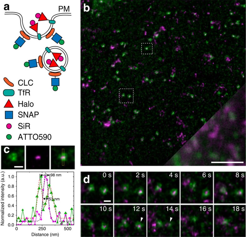
Stimulated emission depletion (STED) nanoscopy allows observations of subcellular dynamics at the nanoscale. Applications have, however, been severely limited by the lack of a versatile STED-compatible two-colour labelling strategy for intracellular targets in living cells. Here we demonstrate a universal labelling method based on the organic, membrane-permeable dyes SiR and ATTO590 as Halo and SNAP substrates. SiR and ATTO590 constitute the first suitable dye pair for two-colour STED imaging in living cells below 50 nm resolution. We show applications with mitochondria, endoplasmic reticulum, plasma membrane and Golgi-localized proteins, and demonstrate continuous acquisition for up to 3 min at 2-s time resolution.
受激发射损耗(STED)纳米显微镜能够在纳米尺度上观察亚细胞动力学。然而,由于缺乏一种适用于活细胞内靶点的通用的与STED兼容的双色标记策略,其应用受到了严重限制。在此,我们展示了一种基于有机的、可透过细胞膜的染料SiR和ATTO590作为Halo和SNAP底物的通用标记方法。SiR和ATTO590构成了第一对适用于分辨率低于50 nm的活细胞双色STED成像的染料。我们展示了该方法在定位在线粒体、内质网、质膜和高尔基体的蛋白质上的应用,并证明了能够以2秒的时间分辨率连续采集长达3分钟的数据。