Helen Wills Neuroscience Institute, University of California, Berkeley, Berkeley, United States.
Department of Anesthesiology and Intensive Care Medicine, University Medical Center Tuebingen, Tuebingen, Germany.
Elife. 2020 Jul 28;9:e55092. doi: 10.7554/eLife.55092.
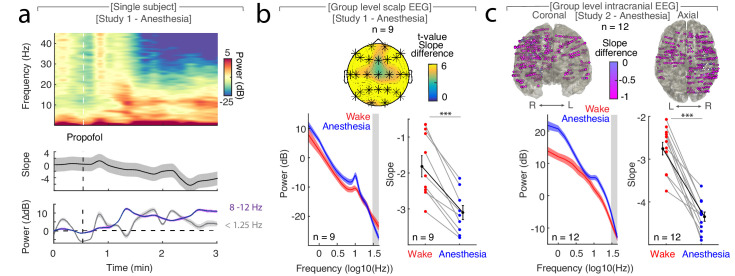
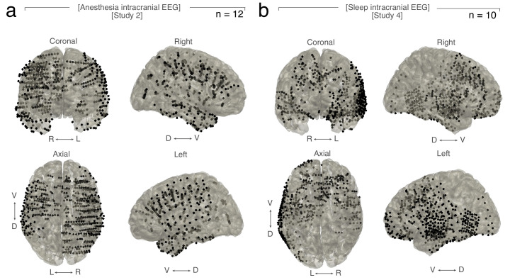
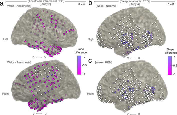
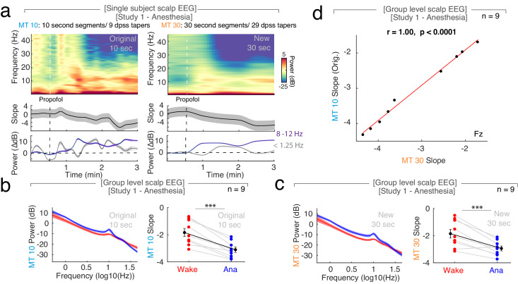
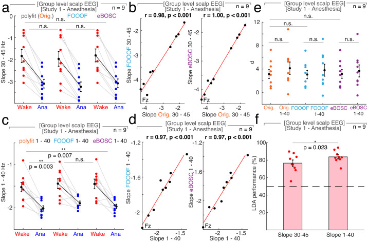
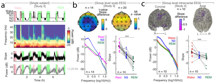
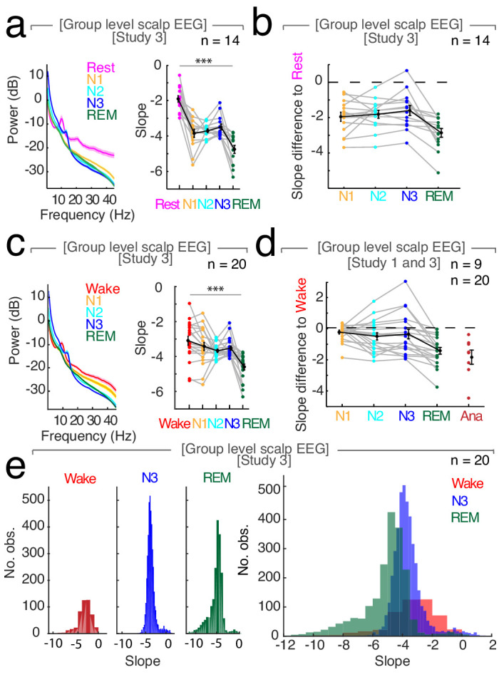
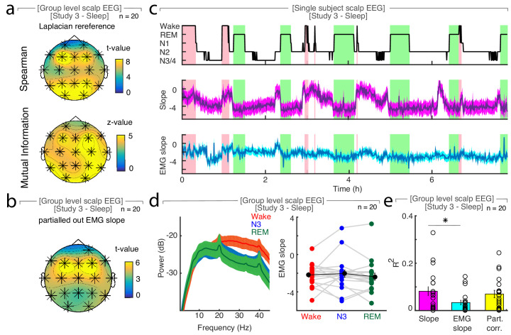
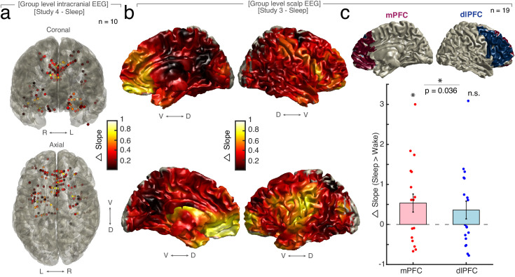
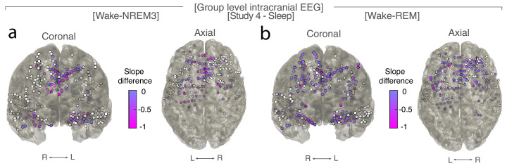
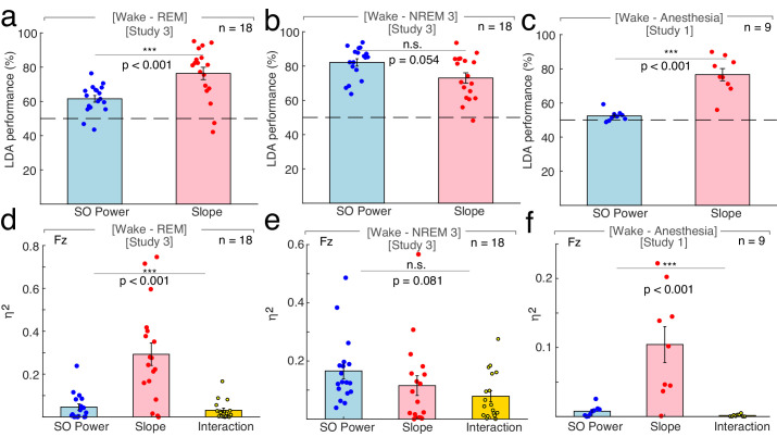
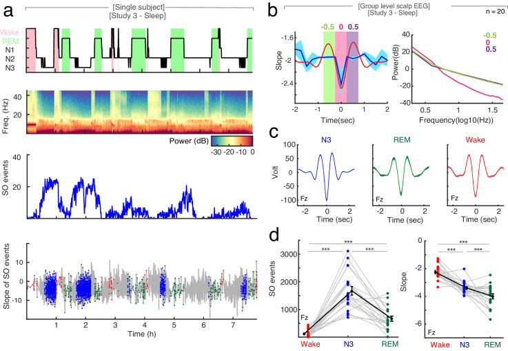
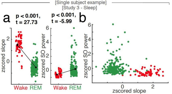
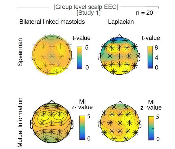
Deep non-rapid eye movement sleep (NREM) and general anesthesia with propofol are prominent states of reduced arousal linked to the occurrence of synchronized oscillations in the electroencephalogram (EEG). Although rapid eye movement (REM) sleep is also associated with diminished arousal levels, it is characterized by a desynchronized, 'wake-like' EEG. This observation implies that reduced arousal states are not necessarily only defined by synchronous oscillatory activity. Using intracranial and surface EEG recordings in four independent data sets, we demonstrate that the 1/f spectral slope of the electrophysiological power spectrum, which reflects the non-oscillatory, scale-free component of neural activity, delineates wakefulness from propofol anesthesia, NREM and REM sleep. Critically, the spectral slope discriminates wakefulness from REM sleep solely based on the neurophysiological brain state. Taken together, our findings describe a common electrophysiological marker that tracks states of reduced arousal, including different sleep stages as well as anesthesia in humans.
深度非快速眼动睡眠(NREM)和异丙酚全身麻醉是降低觉醒状态的突出表现,与脑电图(EEG)中的同步振荡有关。尽管快速眼动(REM)睡眠也与觉醒水平降低有关,但它的特点是去同步化,表现为“清醒样”脑电图。这一观察结果表明,降低觉醒状态不一定仅由同步振荡活动来定义。我们使用四个独立数据集的颅内和表面 EEG 记录,证明了电生理功率谱的 1/f 频谱斜率,它反映了神经活动的非振荡、无标度成分,将清醒状态与异丙酚麻醉、NREM 和 REM 睡眠区分开来。关键的是,频谱斜率仅根据神经生理脑状态将清醒状态与 REM 睡眠区分开来。总之,我们的发现描述了一个共同的电生理标记,它可以跟踪降低觉醒状态,包括不同的睡眠阶段以及人类的麻醉状态。