Charalampaki Patra, Proskynitopoulos Phileas Johannes, Heimann Axel, Nakamura Makoto
Department of Neurosurgery, Cologne Medical Center, Cologne, Germany.
Witten-Herdecke University, Witten, Germany.
Front Oncol. 2020 Jul 8;10:1069. doi: 10.3389/fonc.2020.01069. eCollection 2020.
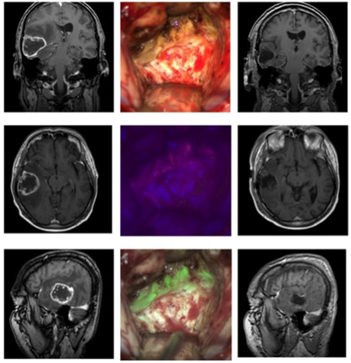
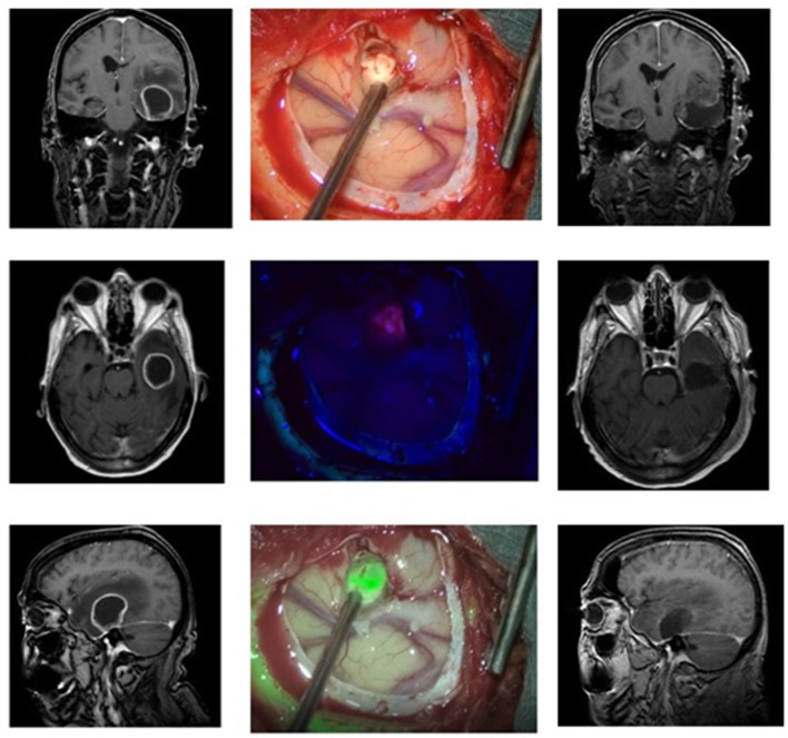
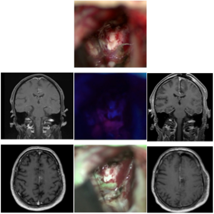
Fluorescence-guided surgery with five-aminolevulinic acid (5-ALA) is the state-of-the-art treatment of high-grade gliomas. However, intraoperative visualization of 5-ALA under blue light remains challenging, especially when blood covers the surgical field and thereby fluorescence. To overcome this problem and combine the brightness of visible light with the information delivered with fluorescence, we implemented multispectral fluorescence (MFL) in a surgical microscope, a technique that is able to project both information in real-time. We prospectively examined 25 patients with brain tumors. One patient was operated on two different lesions in the same setting. The tumors comprised: six glioblastomas, four anaplastic astrocytomas, one anaplastic oligodendroglioma, two meningiomas, 11 metastatic tumors, one acoustic neuroma, and one ependymoma. The MFL technique with a real-time overlay of fluorescence and white light was compared intraoperatively to the classic blue filter. All lesions were clearly visible and highlighted from the surrounding tissue. The pseudocolor we chose was green, representing fluorescence, with the surrounding brain tissue remaining in its original color. When blood was covering the surgical field, orientation was easy to maintain. The MFL technique opens the way for precise and clear visualization of fluorescence in real-time under white light. It can be easily used for the resection of all tumors accumulating 5-ALA. Drawbacks of classic PpIX fluorescence such as hidden fluorescence, intraoperative changes could be overcome with the presence of additional white light in MFL technique.
使用5-氨基乙酰丙酸(5-ALA)的荧光引导手术是高级别胶质瘤的先进治疗方法。然而,蓝光下5-ALA的术中可视化仍然具有挑战性,尤其是当血液覆盖手术视野并因此掩盖荧光时。为了克服这个问题并将可见光的亮度与荧光传递的信息相结合,我们在手术显微镜中实现了多光谱荧光(MFL),这是一种能够实时投射两种信息的技术。我们前瞻性地检查了25例脑肿瘤患者。一名患者在同一情况下对两个不同的病变进行了手术。肿瘤包括:6例胶质母细胞瘤、4例间变性星形细胞瘤、1例间变性少突胶质细胞瘤、2例脑膜瘤、11例转移瘤、1例听神经瘤和1例室管膜瘤。术中将具有荧光和白光实时叠加的MFL技术与经典蓝色滤光片进行了比较。所有病变都清晰可见,并与周围组织形成鲜明对比。我们选择的伪彩色是绿色,代表荧光,周围脑组织保持其原始颜色。当血液覆盖手术视野时,很容易保持手术方向。MFL技术为在白光下实时精确清晰地可视化荧光开辟了道路。它可以很容易地用于切除所有积累5-ALA的肿瘤。经典的原卟啉IX(PpIX)荧光的缺点,如隐藏荧光、术中变化,都可以通过MFL技术中额外白光的存在来克服。