• 文献检索
  • 文档翻译
  • 深度研究
  • 学术资讯
  • Suppr Zotero 插件Zotero 插件
  • 邀请有礼
  • 套餐&价格
  • 历史记录
应用&插件
Suppr Zotero 插件Zotero 插件浏览器插件Mac 客户端Windows 客户端微信小程序
定价
高级版会员购买积分包购买API积分包
服务
文献检索文档翻译深度研究API 文档MCP 服务
关于我们
关于 Suppr公司介绍联系我们用户协议隐私条款
关注我们

Suppr 超能文献

核心技术专利:CN118964589B侵权必究
粤ICP备2023148730 号-1Suppr @ 2026

文献检索

告别复杂PubMed语法,用中文像聊天一样搜索,搜遍4000万医学文献。AI智能推荐,让科研检索更轻松。

立即免费搜索

文件翻译

保留排版,准确专业,支持PDF/Word/PPT等文件格式,支持 12+语言互译。

免费翻译文档

深度研究

AI帮你快速写综述,25分钟生成高质量综述,智能提取关键信息,辅助科研写作。

立即免费体验

利用目标识别实现前列腺 T2 加权磁共振图像的自动参考组织标准化。

Automated reference tissue normalization of T2-weighted MR images of the prostate using object recognition.

机构信息

Department of Circulation and Medical Imaging, NTNU, Norwegian University of Science and Technology, 7030, Trondheim, Norway.

Department of Radiology and Nuclear Medicine, St. Olavs Hospital, Trondheim University Hospital, 7030, Trondheim, Norway.

出版信息

MAGMA. 2021 Apr;34(2):309-321. doi: 10.1007/s10334-020-00871-3. Epub 2020 Jul 31.

DOI:10.1007/s10334-020-00871-3
PMID:32737628
原文链接:https://pmc.ncbi.nlm.nih.gov/articles/PMC8018925/
Abstract

OBJECTIVES

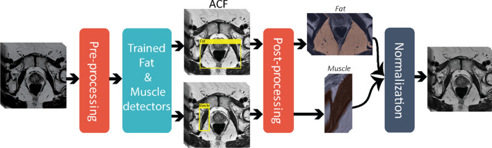
To develop and evaluate an automated method for prostate T2-weighted (T2W) image normalization using dual-reference (fat and muscle) tissue.

MATERIALS AND METHODS

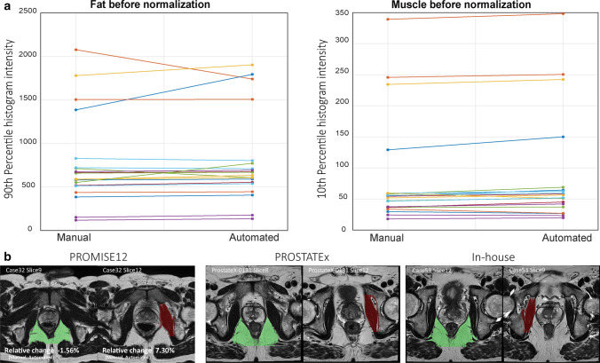
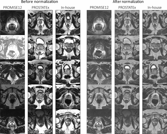
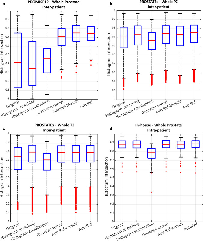
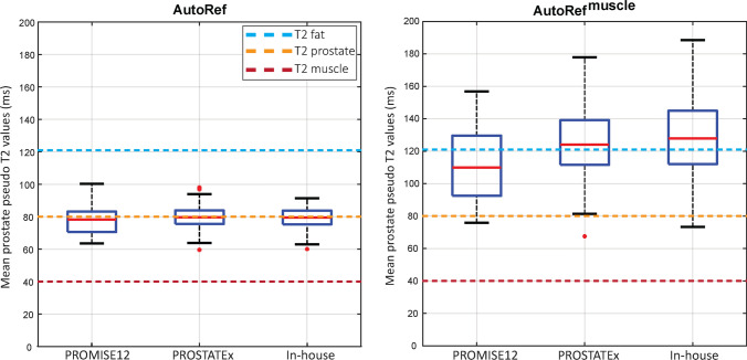
Transverse T2W images from the publicly available PROMISE12 (N = 80) and PROSTATEx (N = 202) challenge datasets, and an in-house collected dataset (N = 60) were used. Aggregate channel features object detectors were trained to detect reference fat and muscle tissue regions, which were processed and utilized to normalize the 3D images by linear scaling. Mean prostate pseudo T2 values after normalization were compared to literature values. Inter-patient histogram intersections of voxel intensities in the prostate were compared between our approach, the original images, and other commonly used normalization methods. Healthy vs. malignant tissue classification performance was compared before and after normalization.

RESULTS

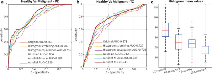
The prostate pseudo T2 values of the three tested datasets (mean ± standard deviation = 78.49 ± 9.42, 79.69 ± 6.34 and 79.29 ± 6.30 ms) corresponded well to T2 values from literature (80 ± 34 ms). Our normalization approach resulted in significantly higher (p < 0.001) inter-patient histogram intersections (median = 0.746) than the original images (median = 0.417) and most other normalization methods. Healthy vs. malignant classification also improved significantly (p < 0.001) in peripheral (AUC 0.826 vs. 0.769) and transition (AUC 0.743 vs. 0.678) zones.

CONCLUSION

An automated dual-reference tissue normalization of T2W images could help improve the quantitative assessment of prostate cancer.

摘要

目的

开发并评估一种使用双参考(脂肪和肌肉)组织对前列腺 T2 加权(T2W)图像进行归一化的自动方法。

材料与方法

使用了来自公开的 PROMISE12(N=80)和 PROSTATEx(N=202)挑战赛数据集以及内部收集的数据集(N=60)的横向 T2W 图像。聚合通道特征目标检测器被训练来检测参考脂肪和肌肉组织区域,这些区域经过处理并用于通过线性缩放来归一化 3D 图像。归一化后前列腺的平均伪 T2 值与文献值进行了比较。比较了我们的方法、原始图像和其他常用归一化方法在前列腺中体素强度的患者间直方图交点。比较了归一化前后健康与恶性组织的分类性能。

结果

三个测试数据集(平均±标准差=78.49±9.42、79.69±6.34 和 79.29±6.30 ms)的前列腺伪 T2 值与文献中的 T2 值(80±34 ms)非常吻合。与原始图像(中位数=0.417)和大多数其他归一化方法相比,我们的归一化方法导致的患者间直方图交点(中位数=0.746)显著更高(p<0.001)。外周(AUC 0.826 与 0.769)和过渡(AUC 0.743 与 0.678)区的健康与恶性分类也显著改善(p<0.001)。

结论

T2W 图像的自动双参考组织归一化可以帮助改善前列腺癌的定量评估。

https://cdn.ncbi.nlm.nih.gov/pmc/blobs/3d7e/8018925/44bf282498dd/10334_2020_871_Fig6_HTML.jpg
https://cdn.ncbi.nlm.nih.gov/pmc/blobs/3d7e/8018925/47108d1b148a/10334_2020_871_Fig1_HTML.jpg
https://cdn.ncbi.nlm.nih.gov/pmc/blobs/3d7e/8018925/b0473e9dbeba/10334_2020_871_Fig2_HTML.jpg
https://cdn.ncbi.nlm.nih.gov/pmc/blobs/3d7e/8018925/861a73cfc1cc/10334_2020_871_Fig3_HTML.jpg
https://cdn.ncbi.nlm.nih.gov/pmc/blobs/3d7e/8018925/df8cd4b0a09a/10334_2020_871_Fig4_HTML.jpg
https://cdn.ncbi.nlm.nih.gov/pmc/blobs/3d7e/8018925/0bc17a0ff30c/10334_2020_871_Fig5_HTML.jpg
https://cdn.ncbi.nlm.nih.gov/pmc/blobs/3d7e/8018925/44bf282498dd/10334_2020_871_Fig6_HTML.jpg

相似文献

1
Automated reference tissue normalization of T2-weighted MR images of the prostate using object recognition.利用目标识别实现前列腺 T2 加权磁共振图像的自动参考组织标准化。
MAGMA. 2021 Apr;34(2):309-321. doi: 10.1007/s10334-020-00871-3. Epub 2020 Jul 31.
2
Pseudo-T2 mapping for normalization of T2-weighted prostate MRI.基于假性 T2 映射的前列腺 MRI T2 加权图像标准化。
MAGMA. 2022 Aug;35(4):573-585. doi: 10.1007/s10334-022-01003-9. Epub 2022 Feb 12.
3
Uncovering prostate cancer aggressiveness signal in T2-weighted MRI through a three-reference tissues normalization technique.通过一种三参照组织归一化技术揭示 T2 加权 MRI 中的前列腺癌侵袭性信号。
NMR Biomed. 2024 Mar;37(3):e5069. doi: 10.1002/nbm.5069. Epub 2023 Nov 21.
4
Automated separation of diffusely abnormal white matter from focal white matter lesions on MRI in multiple sclerosis.在多发性硬化症的 MRI 上自动分离弥漫性异常的白质和局灶性白质病变。
Neuroimage. 2020 Jun;213:116690. doi: 10.1016/j.neuroimage.2020.116690. Epub 2020 Feb 29.
5
T2 Mapping in Prostate Cancer.前列腺癌的 T2 映射。
Invest Radiol. 2019 Mar;54(3):146-152. doi: 10.1097/RLI.0000000000000520.
6
Radiomics based automated quality assessment for T2W prostate MR images.基于放射组学的 T2W 前列腺磁共振图像自动质量评估。
Eur J Radiol. 2023 Aug;165:110928. doi: 10.1016/j.ejrad.2023.110928. Epub 2023 Jun 15.
7
A dual model HU conversion from MRI intensity values within and outside of bone segment for MRI-based radiotherapy treatment planning of prostate cancer.一种用于基于MRI的前列腺癌放射治疗计划的双模型HU转换,该转换基于骨段内外的MRI强度值。
Med Phys. 2014 Jan;41(1):011704. doi: 10.1118/1.4842575.
8
Automated segmentation of prostate zonal anatomy on T2-weighted (T2W) and apparent diffusion coefficient (ADC) map MR images using U-Nets.基于 U-Nets 的 T2 加权(T2W)和表观扩散系数(ADC)图磁共振成像上前列腺分区解剖结构的自动分割。
Med Phys. 2019 Jul;46(7):3078-3090. doi: 10.1002/mp.13550. Epub 2019 May 11.
9
Is there an additional value of ¹¹C-choline PET-CT to T2-weighted MRI images in the localization of intraprostatic tumor nodules?¹¹C-胆碱 PET-CT 对 T2 加权 MRI 图像在前列腺内肿瘤结节定位中的附加价值如何?
Int J Radiat Oncol Biol Phys. 2012 Aug 1;83(5):1486-92. doi: 10.1016/j.ijrobp.2011.10.046. Epub 2012 Jan 26.
10
Effects of MRI image normalization techniques in prostate cancer radiomics.MRI 图像标准化技术对前列腺癌放射组学的影响。
Phys Med. 2020 Mar;71:7-13. doi: 10.1016/j.ejmp.2020.02.007. Epub 2020 Feb 18.

引用本文的文献

1
A Narrative Review of Artificial Intelligence in MRI-Guided Prostate Cancer Diagnosis: Addressing Key Challenges.磁共振成像引导下前列腺癌诊断中人工智能的叙事性综述:应对关键挑战
Diagnostics (Basel). 2025 May 26;15(11):1342. doi: 10.3390/diagnostics15111342.
2
Comparison of intensity normalization methods in prostate, brain, and breast cancer multi-parametric magnetic resonance imaging.前列腺、脑和乳腺癌多参数磁共振成像中强度归一化方法的比较
Front Oncol. 2025 Feb 7;15:1433444. doi: 10.3389/fonc.2025.1433444. eCollection 2025.
3
Retrospectively Quantified T2 Improves Detection of Clinically Significant Peripheral Zone Prostate Cancer.

本文引用的文献

1
Repeatability of Multiparametric Prostate MRI Radiomics Features.多参数前列腺 MRI 放射组学特征的可重复性。
Sci Rep. 2019 Jul 1;9(1):9441. doi: 10.1038/s41598-019-45766-z.
2
PROSTATEx Challenges for computerized classification of prostate lesions from multiparametric magnetic resonance images.前列腺X:多参数磁共振图像中前列腺病变的计算机分类面临的挑战
J Med Imaging (Bellingham). 2018 Oct;5(4):044501. doi: 10.1117/1.JMI.5.4.044501. Epub 2018 Nov 10.
3
Global cancer statistics 2018: GLOBOCAN estimates of incidence and mortality worldwide for 36 cancers in 185 countries.
回顾性定量T2加权成像可提高临床显著外周带前列腺癌的检出率。
Cancers (Basel). 2025 Jan 24;17(3):381. doi: 10.3390/cancers17030381.
4
Integrated framework for quantitative T2-weighted MRI analysis following prostate cancer radiotherapy.前列腺癌放疗后定量T2加权磁共振成像分析的综合框架
Phys Imaging Radiat Oncol. 2024 Oct 24;32:100660. doi: 10.1016/j.phro.2024.100660. eCollection 2024 Oct.
5
Magnetic resonance imaging preprocessing and radiomic features for classification of autosomal dominant polycystic kidney disease genotype.用于常染色体显性多囊肾病基因型分类的磁共振成像预处理及影像组学特征
J Med Imaging (Bellingham). 2023 Nov;10(6):064503. doi: 10.1117/1.JMI.10.6.064503. Epub 2023 Dec 27.
6
The impact of pre-processing and disease characteristics on reproducibility of T2-weighted MRI radiomics features.预处理和疾病特征对 T2 加权 MRI 放射组学特征可重复性的影响。
MAGMA. 2023 Dec;36(6):945-956. doi: 10.1007/s10334-023-01112-z. Epub 2023 Aug 9.
7
CROPro: a tool for automated cropping of prostate magnetic resonance images.CROPro:一种用于自动裁剪前列腺磁共振图像的工具。
J Med Imaging (Bellingham). 2023 Mar;10(2):024004. doi: 10.1117/1.JMI.10.2.024004. Epub 2023 Mar 7.
8
A Framework of Analysis to Facilitate the Harmonization of Multicenter Radiomic Features in Prostate Cancer.促进前列腺癌多中心放射组学特征协调统一的分析框架
J Clin Med. 2022 Dec 24;12(1):140. doi: 10.3390/jcm12010140.
9
Artificial intelligence for prostate MRI: open datasets, available applications, and grand challenges.前列腺 MRI 的人工智能:开放数据集、现有应用和重大挑战。
Eur Radiol Exp. 2022 Aug 1;6(1):35. doi: 10.1186/s41747-022-00288-8.
10
A New Framework for Precise Identification of Prostatic Adenocarcinoma.一种前列腺腺癌精准识别的新框架。
Sensors (Basel). 2022 Feb 26;22(5):1848. doi: 10.3390/s22051848.
全球癌症统计数据 2018:GLOBOCAN 对全球 185 个国家/地区 36 种癌症的发病率和死亡率的估计。
CA Cancer J Clin. 2018 Nov;68(6):394-424. doi: 10.3322/caac.21492. Epub 2018 Sep 12.
4
Prostate cancer radiomics and the promise of radiogenomics.前列腺癌影像组学与放射基因组学的前景
Transl Cancer Res. 2016 Aug;5(4):432-447. doi: 10.21037/tcr.2016.06.20.
5
A survey on deep learning in medical image analysis.深度学习在医学图像分析中的应用研究综述。
Med Image Anal. 2017 Dec;42:60-88. doi: 10.1016/j.media.2017.07.005. Epub 2017 Jul 26.
6
A novel alternative to classify tissues from T 1 and T 2 relaxation times for prostate MRI.一种用于前列腺MRI中根据T1和T2弛豫时间对组织进行分类的新型替代方法。
MAGMA. 2016 Oct;29(5):777-88. doi: 10.1007/s10334-016-0562-3. Epub 2016 May 9.
7
PI-RADS Prostate Imaging - Reporting and Data System: 2015, Version 2.PI-RADS前列腺影像报告和数据系统:2015版,第2版
Eur Urol. 2016 Jan;69(1):16-40. doi: 10.1016/j.eururo.2015.08.052. Epub 2015 Oct 1.
8
Fast Feature Pyramids for Object Detection.快速目标检测特征金字塔。
IEEE Trans Pattern Anal Mach Intell. 2014 Aug;36(8):1532-45. doi: 10.1109/TPAMI.2014.2300479.
9
Zone-specific logistic regression models improve classification of prostate cancer on multi-parametric MRI.区域特异性逻辑回归模型可改善多参数磁共振成像对前列腺癌的分类。
Eur Radiol. 2015 Sep;25(9):2727-37. doi: 10.1007/s00330-015-3636-0. Epub 2015 Feb 14.
10
Computer aided-diagnosis of prostate cancer on multiparametric MRI: a technical review of current research.多参数磁共振成像上前列腺癌的计算机辅助诊断:当前研究的技术综述
Biomed Res Int. 2014;2014:789561. doi: 10.1155/2014/789561. Epub 2014 Dec 1.