Mehanna Ahmed Mohamed, Abdelnaby Moustafa Mohamed, Eid Mohamed
Department of Otolaryngology, Alexandria University, Midan al Khartoum, Alexandria, Egypt.
Department of Radiodiagnosis, Faculty of Medicine of Alexandria University, Midan al Khartoum, Alexandria, Egypt.
Int Arch Otorhinolaryngol. 2020 Jul;24(3):e288-e298. doi: 10.1055/s-0039-1698783. Epub 2019 Dec 13.
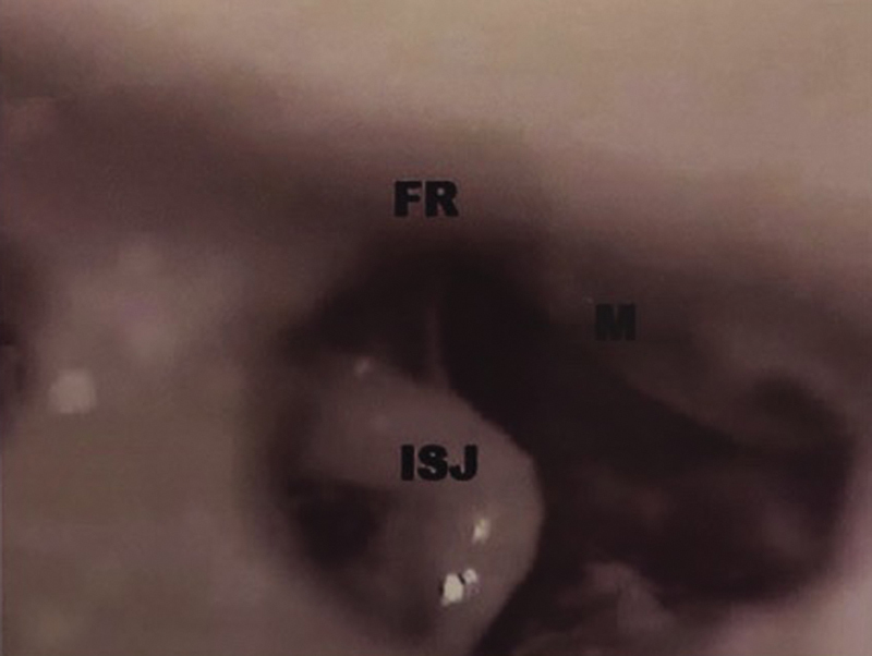
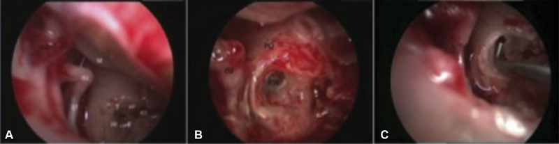
Over the last decades, there has been a tremendous increase in the number of cochlear implant recipients and, consequently, there is a recent increase of interest in the proper understanding of the anatomy of the round window (RW), which is the most important anatomical land mark during cochlear implant surgery. The present study was undertaken to assess the detailed surgical and radiological anatomy of the RW prechamber; its shape, directions, measurements, common anatomic variations, and its relationships with different surrounding structures as related to cochlear implantation. A total of 20 cadaveric specimens of human temporal bone were microscopically dissected for the anatomical assessment of the measurements of the RW and its relation to surrounding structures in the tympanum. A total of 20 patients were subjected to cochlear implantation, and a radiological and surgical assessment of the anatomy of their RW prechambers was performed. The distances between the RW and the facial canal (FC), the jugular fossa (JF), the carotid canal (CC), and the oval window (OW) were measured. Among the cases subjected to cochlear implantation, the infracochlear tunnel was studied radiologically; the lengths of the anterior and posterior pillars were assessed, and the relation with the direction at which the RW faces was statistically analyzed. Proper understanding of the topographic anatomy of the RW, including its direction of opening and the distances from different adjacent structures in the tympanum, is essential for a successful cochlear implantation surgery, since it can help decision-making before the surgery and is useful to avoid many complications, such as misplaced electrode and iatrogenic injury to the surrounding structures.
在过去几十年中,人工耳蜗植入受者的数量大幅增加,因此,最近人们对正确理解圆窗(RW)的解剖结构越来越感兴趣,圆窗是人工耳蜗植入手术中最重要的解剖标志。本研究旨在评估圆窗前庭的详细手术和放射解剖结构;其形状、方向、测量值、常见解剖变异以及与人工耳蜗植入相关的不同周围结构的关系。对20个人类颞骨尸体标本进行显微镜解剖,以评估圆窗的测量值及其与鼓室周围结构的关系。共有20例患者接受了人工耳蜗植入,并对其圆窗前庭的解剖结构进行了放射学和手术评估。测量了圆窗与面神经管(FC)、颈静脉窝(JF)、颈动脉管(CC)和卵圆窗(OW)之间的距离。在接受人工耳蜗植入的病例中,对耳蜗下隧道进行了放射学研究;评估了前后支柱的长度,并对其与圆窗所朝方向的关系进行了统计分析。正确理解圆窗的局部解剖结构,包括其开口方向和与鼓室内不同相邻结构的距离,对于人工耳蜗植入手术的成功至关重要,因为它有助于术前决策,并有助于避免许多并发症,如电极放置不当和对周围结构的医源性损伤。