Graduate School of Information Science and Technology, Hokkaido University, Sapporo, Hokkaido, Japan.
Research Institute for Electronic Science, Hokkaido University, Sapporo, Hokkaido, Japan.
PLoS One. 2020 Aug 7;15(8):e0237230. doi: 10.1371/journal.pone.0237230. eCollection 2020.
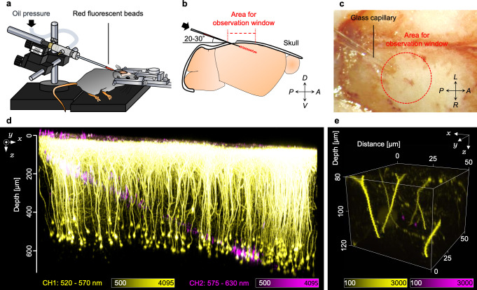
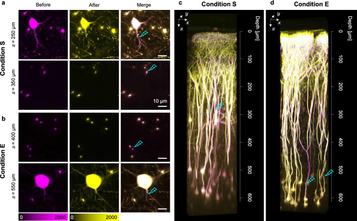
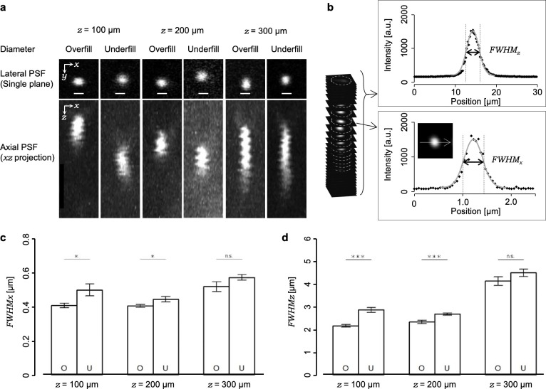
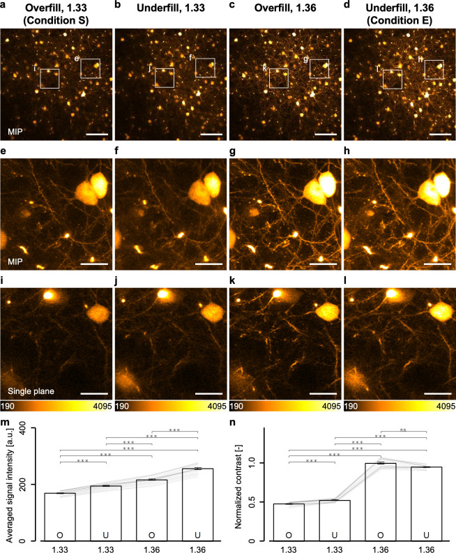
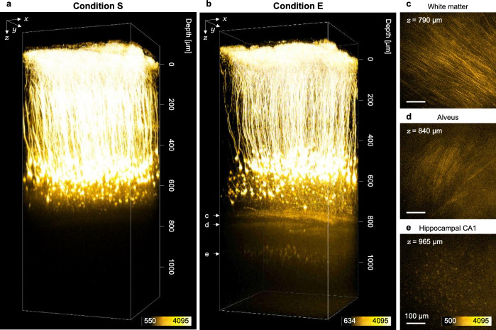
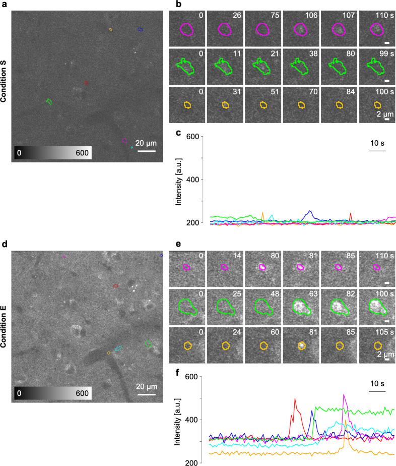
In vivo two-photon microscopy utilizing a nonlinear optical process enables, in living mouse brains, not only the visualization of morphologies and functions of neural networks in deep regions but also their optical manipulation at targeted sites with high spatial precision. Because the two-photon excitation efficiency is proportional to the square of the photon density of the excitation laser light at the focal position, optical aberrations induced by specimens mainly limit the maximum depth of observations or that of manipulations in the microscopy. To increase the two-photon excitation efficiency, we developed a method for evaluating the focal volume in living mouse brains. With this method, we modified the beam diameter of the excitation laser light and the value of the refractive index in the immersion liquid to maximize the excitation photon density at the focal position. These two modifications allowed the successful visualization of the finer structures of hippocampal CA1 neurons, as well as the intracellular calcium dynamics in cortical layer V astrocytes, even with our conventional two-photon microscopy system. Furthermore, it enabled focal laser ablation dissection of both single apical and single basal dendrites of cortical layer V pyramidal neurons. These simple modifications would enable us to investigate the contributions of single cells or single dendrites to the functions of local cortical networks.
利用非线性光学过程的体内双光子显微镜技术,不仅能够在活体小鼠大脑中可视化深层区域神经网络的形态和功能,还能够在靶向部位进行高空间精度的光学操纵。由于双光子激发效率与激发激光在焦平面上的光密度的平方成正比,因此样本引起的光学像差主要限制了显微镜观察或操作的最大深度。为了提高双光子激发效率,我们开发了一种评估活体小鼠大脑中焦体积的方法。通过这种方法,我们改变了激发激光的光束直径和浸液中的折射率,以最大化焦平面上的激发光密度。这两种改进使得我们能够成功地可视化海马 CA1 神经元的更精细结构,以及皮质层 V 星形胶质细胞内的钙动力学,即使使用我们的传统双光子显微镜系统也是如此。此外,它还能够对皮质层 V 锥体神经元的单个顶树突和单个基底树突进行聚焦激光消融分离。这些简单的改进将使我们能够研究单个细胞或单个树突对局部皮质网络功能的贡献。