Lechner Johann, Mayer Wolfgang
Immunology, Clinic Integrative Dentistry, Munich, Germany.
Laboratory, Lab4more, Munich, Germany.
Int J Gen Med. 2020 Jul 10;13:387-402. doi: 10.2147/IJGM.S258170. eCollection 2020.
Mitochondriopathy has recently been linked to several immune system diseases. Historically, there have been many conversations regarding the possible toxic effects of root-filled teeth (RFT), although discussions about the possible decreases in adenosine triphosphate (ATP) activity on the mitochondrial membrane, as caused by dental toxins, are rare. In fact, only a few methods currently exist to assess toxin release in teeth.
An experimental clinical study design is used to investigate the extent to which RFT release toxins in a solution created specifically following extraction (Tox-sol). Our laboratory is investigating the extent to which these Tox-sols reduce ATP activity in patients.
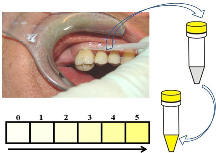
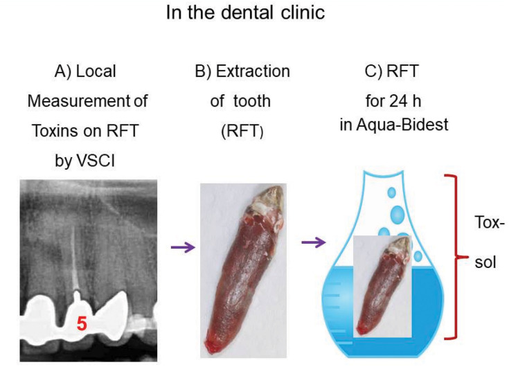
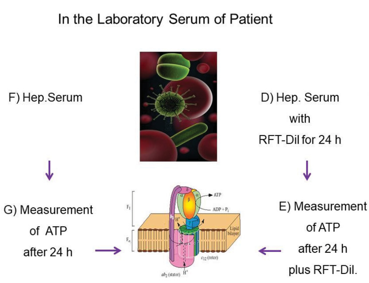
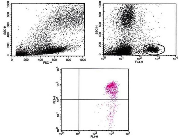
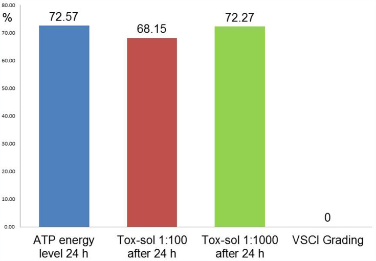
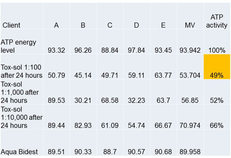
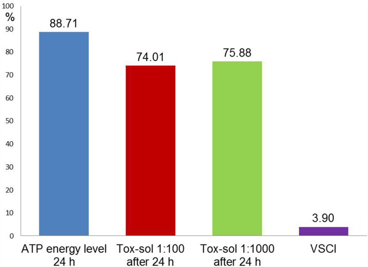
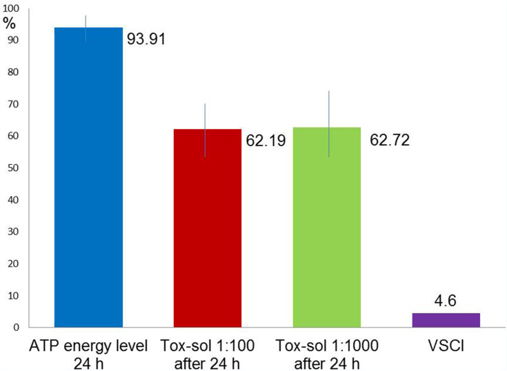
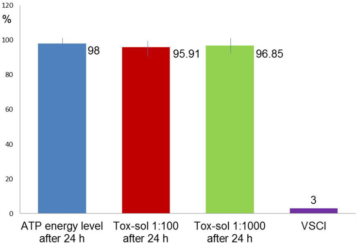
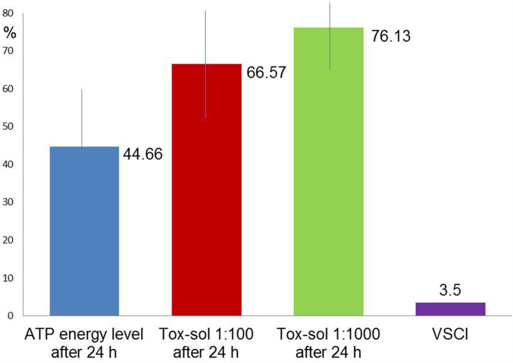
RFTs were identified and extracted to assess their local toxin release using a semi-quantitative volatile sulfur compound indicator (VSCI). These RFTs are placed in an aqueous solution at room temperature for 24 hours and subsequently removed. The resulting solution (Tox-sol) is diluted to 1:100; peripheral blood mononuclear cells (PBMCs) obtained from patients were added to the solution in the laboratory. The remaining ATP activity was measured on the mitochondrial membrane and was compared with the baseline ATP activity of each patient.
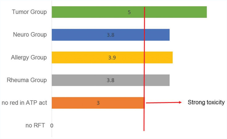
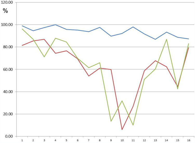
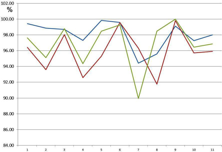
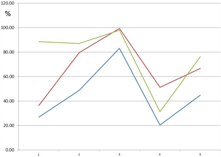
The total population (n=30) showed a ~10% reduction in ATP activity following 24 hours of exposure to the Tox-sol. Three groups emerged with greatly reduced (n=16), neutral (n=10), and increased (n=4) ATP activity. In four different disease groups (rheumatism, neurological disorders, allergies, and tumors), a non-disease specific inhibition of ATP activity was observed.
The study design was limited, as patients were exposed to the Tox-sol and PBMC fraction for only 24 hours. The actual exposure time in a patient's mouth can continue for years and the actual levels can increase over time. Disease-specific effects of Tox-sol were not found.
Within the short exposure time of 24 hours, and at a dilution of 1:100, the Tox-sol caused a median decrease in ATP activity of ~15% in 50% of test subjects. A practical VSCI reliably showed the effects of toxic sulfur compounds on the RFT. The toxic degradation products of biogenic amines from RFT can thus serve as possible contributing factors in the development of mitochondriopathies.
线粒体病最近被认为与多种免疫系统疾病有关。从历史上看,关于根管充填牙(RFT)可能的毒性作用有过很多讨论,不过关于牙科毒素导致线粒体膜上三磷酸腺苷(ATP)活性可能降低的讨论却很少。事实上,目前仅有少数几种方法可用于评估牙齿中的毒素释放情况。
采用实验性临床研究设计,调查RFT在拔牙后专门配制的溶液(毒素溶液)中释放毒素的程度。我们实验室正在研究这些毒素溶液在多大程度上会降低患者的ATP活性。
识别并拔除RFT,使用半定量挥发性硫化合物指示剂(VSCI)评估其局部毒素释放情况。将这些RFT在室温下置于水溶液中24小时,随后取出。将所得溶液(毒素溶液)稀释至1:100;在实验室中将从患者获取的外周血单个核细胞(PBMC)加入该溶液中。测量线粒体膜上剩余的ATP活性,并与每位患者的基线ATP活性进行比较。
总样本量(n = 30)在接触毒素溶液24小时后,ATP活性降低了约10%。出现了三组ATP活性大幅降低(n = 16)、无变化(n = 10)和升高(n = 4)的情况。在四个不同疾病组(风湿病、神经疾病、过敏和肿瘤)中,观察到了对ATP活性的非疾病特异性抑制。
该研究设计存在局限性,因为患者仅接触毒素溶液和PBMC组分24小时。在患者口腔中的实际接触时间可能持续数年,且实际水平会随时间增加。未发现毒素溶液的疾病特异性影响。
在24小时的短接触时间内,以1:100的稀释度,毒素溶液在50%的受试对象中使ATP活性中位数降低了约15%。一种实用的VSCI可靠地显示了有毒硫化合物对RFT的影响。因此,RFT中生物胺的有毒降解产物可能是线粒体病发生发展的潜在促成因素。