Department of Pharmacology, University of Texas Southwestern Medical Center, Dallas, United States.
Department of Biophysics, University of Texas Southwestern Medical Center, Dallas, United States.
Elife. 2020 Aug 10;9:e58157. doi: 10.7554/eLife.58157.
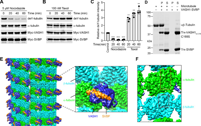
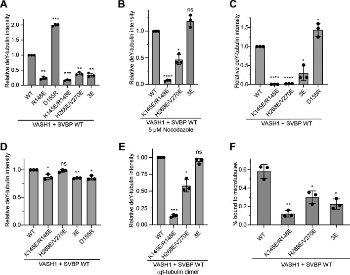
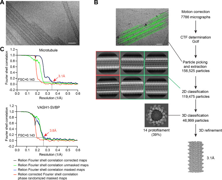
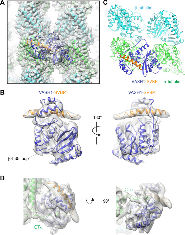
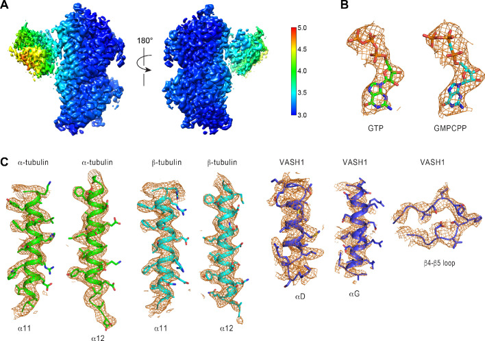
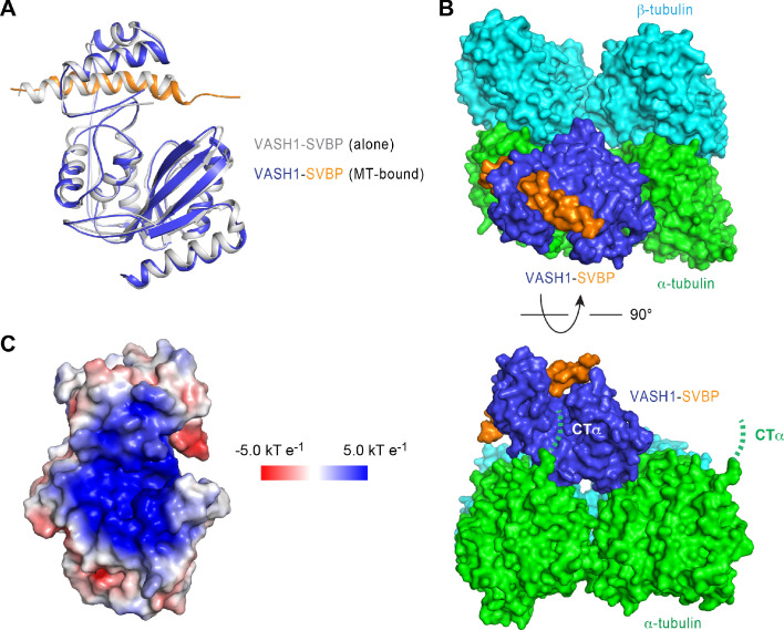
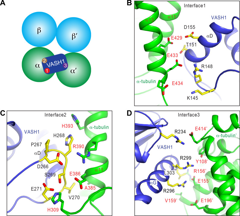
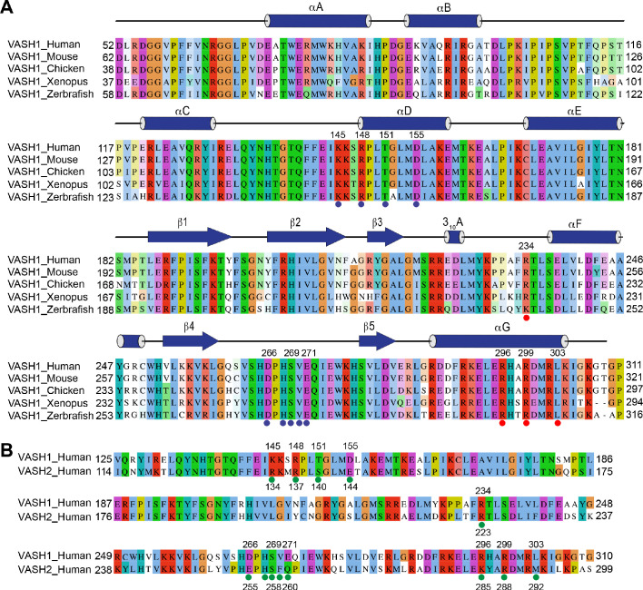
The dynamic tyrosination-detyrosination cycle of α-tubulin regulates microtubule functions. Perturbation of this cycle impairs mitosis, neural physiology, and cardiomyocyte contraction. The carboxypeptidases vasohibins 1 and 2 (VASH1 and VASH2), in complex with the small vasohibin-binding protein (SVBP), mediate α-tubulin detyrosination. These enzymes detyrosinate microtubules more efficiently than soluble αβ-tubulin heterodimers. The structural basis for this substrate preference is not understood. Using cryo-electron microscopy (cryo-EM), we have determined the structure of human VASH1-SVBP bound to microtubules. The acidic C-terminal tail of α-tubulin binds to a positively charged groove near the active site of VASH1. VASH1 forms multiple additional contacts with the globular domain of α-tubulin, including contacts with a second α-tubulin in an adjacent protofilament. Simultaneous engagement of two protofilaments by VASH1 can only occur within the microtubule lattice, but not with free αβ heterodimers. These lattice-specific interactions enable preferential detyrosination of microtubules by VASH1.
α-微管蛋白的动态酪氨酰化-去酪氨酰化循环调节微管功能。该循环的破坏会损害有丝分裂、神经生理学和心肌细胞收缩。羧肽酶血管抑制素 1 和 2(VASH1 和 VASH2)与小血管抑制素结合蛋白(SVBP)形成复合物,介导α-微管蛋白去酪氨酰化。这些酶比可溶性αβ-微管蛋白异二聚体更有效地使微管去酪氨酰化。这种底物偏好的结构基础尚不清楚。我们使用冷冻电镜(cryo-EM)确定了人 VASH1-SVBP 与微管结合的结构。α-微管蛋白的酸性 C 末端尾巴与 VASH1 的活性位点附近的正电荷槽结合。VASH1 与α-微管球蛋白的球状结构域形成多个额外的接触,包括与相邻原纤维中的第二个α-微管蛋白的接触。VASH1 同时与两个原纤维结合只能发生在微管晶格内,而不是与游离的αβ 异二聚体结合。这些晶格特异性相互作用使 VASH1 能够优先对微管进行去酪氨酰化。