Sujiono E H, Zabrian D, Dahlan M Y, Amin B D, Agus J
Laboratory of Materials Physics, Department of Physics, Universitas Negeri Makassar, Makassar, 90224, Indonesia.
Department of Mechanical Engineering, Universitas Negeri Makassar, Makassar, 90224, Indonesia.
Heliyon. 2020 Aug 3;6(8):e04568. doi: 10.1016/j.heliyon.2020.e04568. eCollection 2020 Aug.
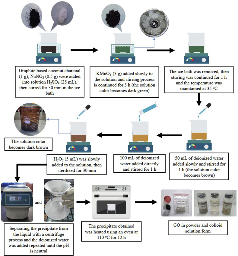
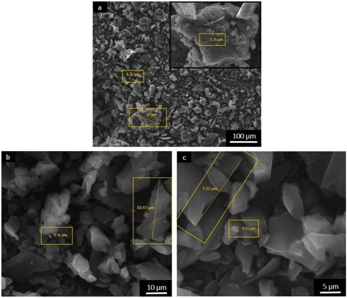
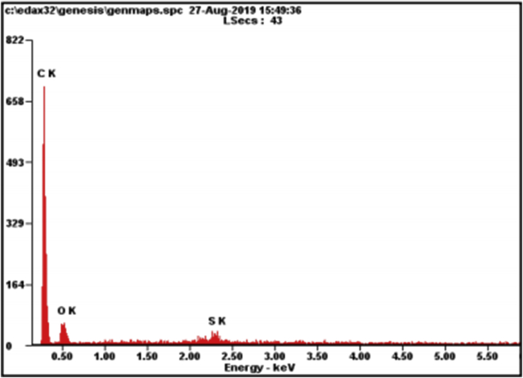
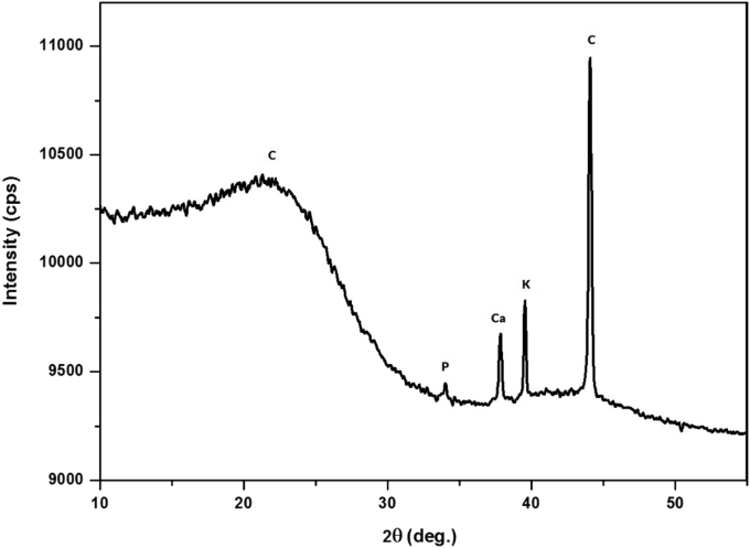
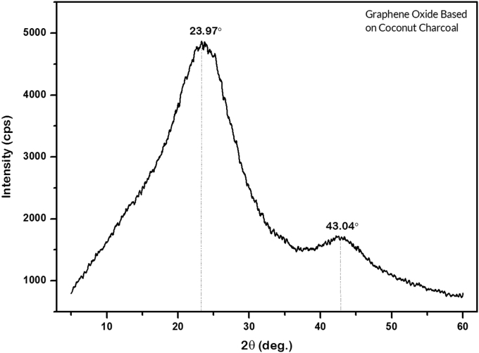
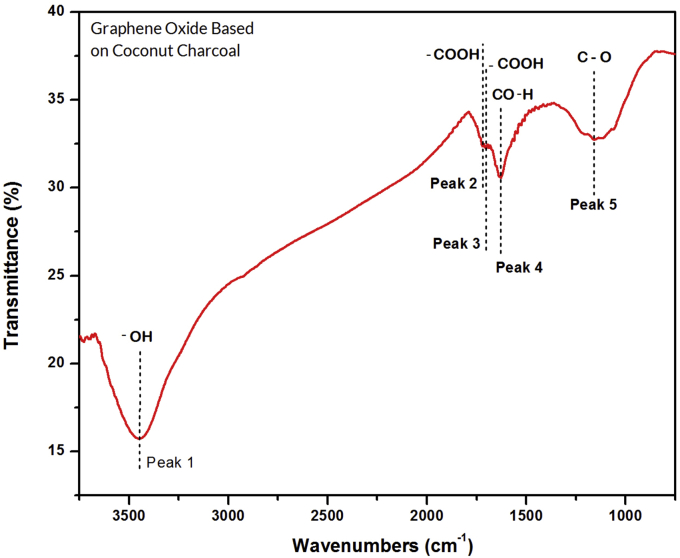
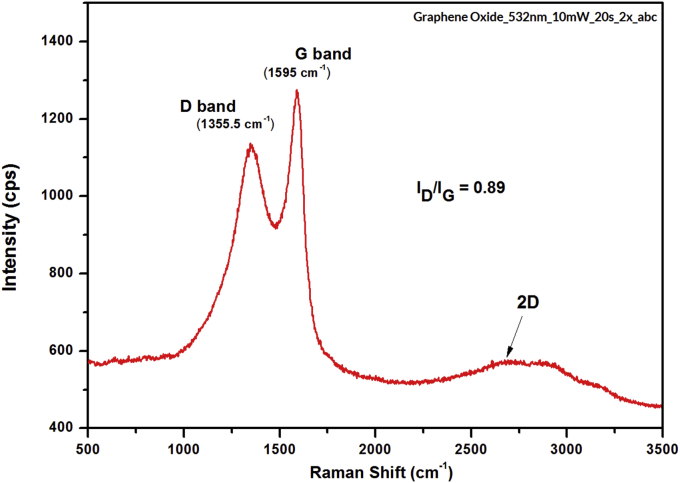
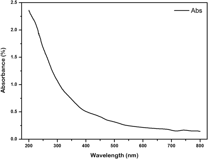
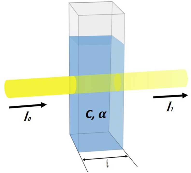
Graphene oxide (GO) based on coconut shell waste was successfully synthesized using a modified Hummers method, and the obtained GO was confirmed using XRD, FTIR, Raman spectroscopy, UV-Vis spectroscopy, and SEM-EDX. The XRD spectroscopy obtained the fractional content of the 2H graphite phase of 71.53%, 14.47% phosphorus, 10.02% calcium, and 3.97% potassium in coconut shell charcoal, where the GO sample tend to forms a phase of reduced graphene oxide (rGO). FTIR spectra shows compound functional groups of hydroxyl (- OH) at peak 1 (3449.92 cm), carboxyl (-COOH) at peak 2 (1719.42 cm) and peak 3 (1702.62 cm), and alcohol (C-OH) at peak 4 (1628.12 cm) and epoxy (CO) at peak 5 (1158.51 cm), which is similar to the GO synthesis from pure graphite. Raman spectroscopy analysis shows that the value of the I/I intensity ratio of the GO sample was 0.89 with a 2D single layer, and SEM results showed that surface morphology with an abundance of granular particles were found with different size distribution. The UV-visible results showed sufficient optical properties characterized by the spectrum, which formed because of the light absorption of the energy passed on the sample. The bandgap energy value of the sample obtained by the Tauc plot method was 4.38 eV, which indicates semiconductor properties.
采用改进的Hummers法成功合成了基于椰壳废料的氧化石墨烯(GO),并通过X射线衍射(XRD)、傅里叶变换红外光谱(FTIR)、拉曼光谱、紫外可见光谱(UV-Vis)和扫描电子显微镜-能谱仪(SEM-EDX)对所得的GO进行了表征。XRD光谱分析得出椰壳炭中2H石墨相的含量为71.53%,磷含量为14.47%,钙含量为10.02%,钾含量为3.97%,其中GO样品倾向于形成还原氧化石墨烯(rGO)相。FTIR光谱显示,在峰1(3449.92 cm)处有羟基(-OH)的复合官能团,在峰2(1719.42 cm)和峰3(1702.62 cm)处有羧基(-COOH),在峰4(1628.12 cm)处有醇基(C-OH),在峰5(1158.51 cm)处有环氧基(CO),这与由纯石墨合成的GO相似。拉曼光谱分析表明,GO样品的I/I强度比为0.89,具有二维单层结构,SEM结果显示表面形态有大量不同尺寸分布的颗粒状颗粒。UV-可见光谱结果表明,样品具有足够的光学性质,这是由于样品对通过的能量的光吸收形成的。通过Tauc图法得到的样品带隙能量值为4.38 eV,表明其具有半导体性质。