Department of Molecular Medicine, The Scripps Research Institute, La Jolla, California, United States.
Eye Center, Medical Center, Faculty of Medicine, University of Freiburg, Freiburg, Germany.
Invest Ophthalmol Vis Sci. 2020 Aug 3;61(10):20. doi: 10.1167/iovs.61.10.20.
Ciliary neurotrophic factor (CNTF) is a well-characterized neurotrophic factor currently in clinical trials for the treatment of macular telangiectasia type II. Our previous work showed that CNTF-induced STAT3 signaling is a potent inhibitor of pathologic preretinal neovascular tuft formation in the mouse model of oxygen-induced retinopathy. In this study, we investigated the effect of CNTF on outer retinal and choroidal angiogenesis and the mechanisms that underpin the observed decrease in outer retinal neovascularization following CNTF treatment.
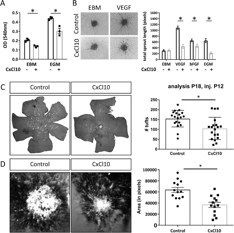
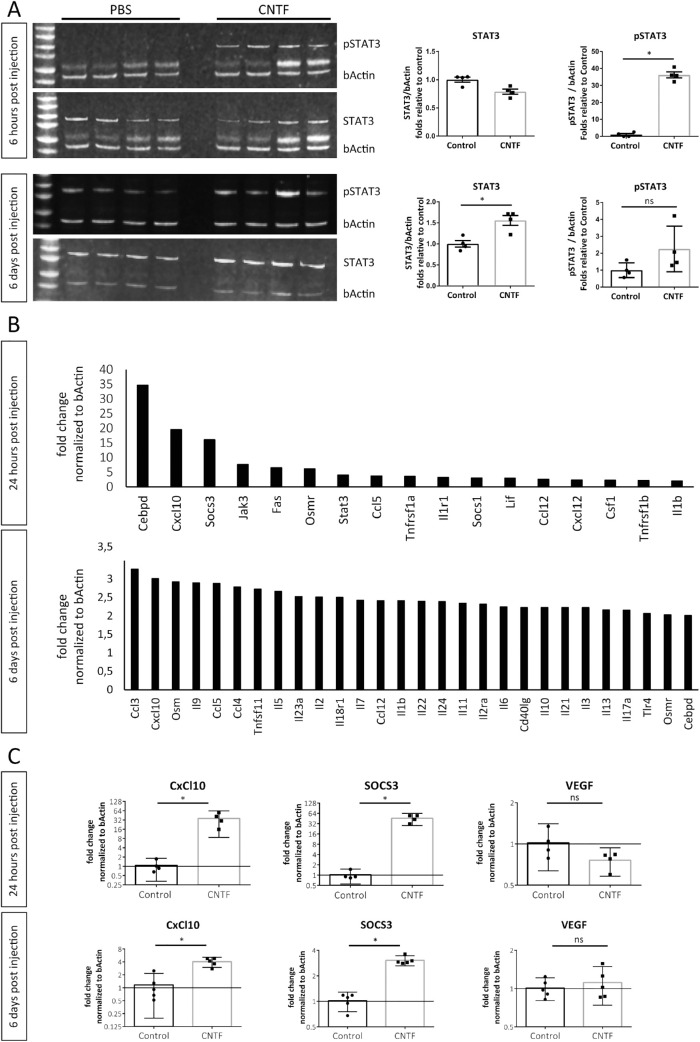
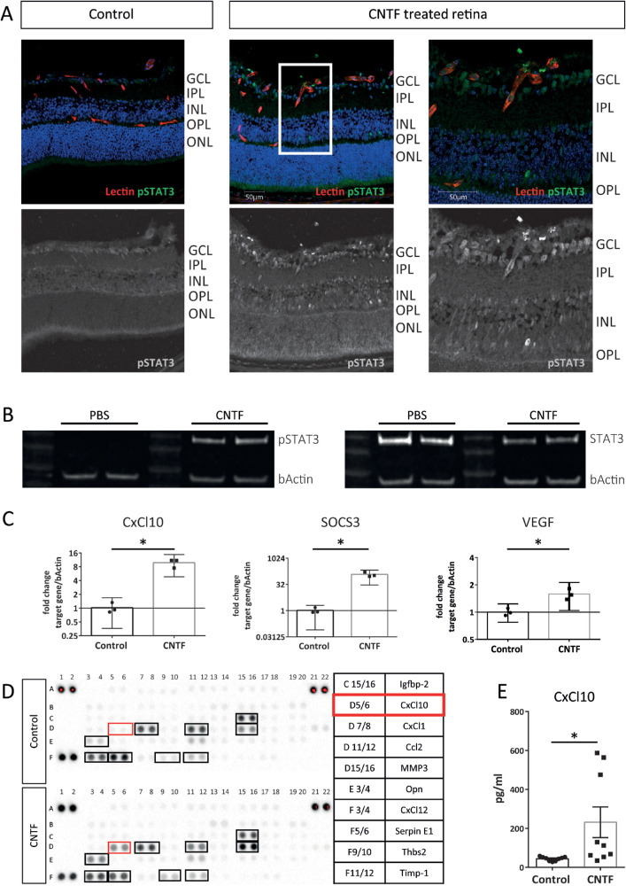
In the Vldlr-/- and laser-CNV mouse models, mice received a one-time injection (on postnatal day [P] 12 in the Vldlr-/- model and 1 day after laser in the Choroidal Neovascularization (CNV) model) of recombinant CNTF or CxCl10, and the extent of neovascular lesions was assessed 6 days posttreatment. STAT3 downstream targets affected by CNTF treatment were identified using quantitative PCR analysis. A proteome array was used to compare media conditioned by CNTF-treated and control-treated primary Müller cells to screen for CNTF-induced changes in secreted angiogenic factors.
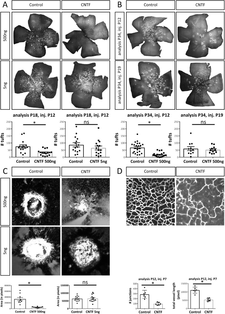
Intravitreal treatment with recombinant CNTF led to significant reduction in neovascularization in the Vldlr-/- and laser-CNV mouse models. Treatment effect in the Vldlr-/- was long-lasting but time sensitive, requiring intravitreal treatment before P19. Mechanistic workup in vitro as well as in vivo confirmed significant activation of the STAT3-signaling pathway in Müller cells in response to CNTF treatment and upregulation of CxCl10. Intravitreal injections of recombinant CxCl10 significantly reduced outer retinal neovascularization in vivo in both the Vldlr-/- and laser-CNV mouse models.
CNTF treatment indirectly affects outer retinal and choroidal neovascularization by inducing CxCl10 secretion from retinal Müller cells.
睫状神经营养因子(CNTF)是一种特征明确的神经营养因子,目前正在进行治疗 II 型黄斑毛细血管扩张症的临床试验。我们之前的工作表明,CNTF 诱导的 STAT3 信号是氧诱导视网膜病变小鼠模型中病理性视网膜前新生血管丛形成的有效抑制剂。在这项研究中,我们研究了 CNTF 对视网膜外层和脉络膜血管生成的影响,以及观察到 CNTF 治疗后视网膜外层新生血管减少的潜在机制。
在 Vldlr-/-和激光-CNV 小鼠模型中,小鼠在出生后第 12 天(Vldlr-/-模型)或激光后第 1 天(脉络膜新生血管(CNV)模型)接受一次重组 CNTF 或 CxCl10 注射,并在治疗后 6 天评估新生血管病变的程度。使用定量 PCR 分析鉴定受 CNTF 处理影响的 STAT3 下游靶标。使用蛋白质组阵列比较 CNTF 处理和对照处理的原代 Müller 细胞的条件培养基,以筛选 CNTF 诱导的分泌血管生成因子的变化。
玻璃体内注射重组 CNTF 可显著减少 Vldlr-/-和激光-CNV 小鼠模型中的新生血管形成。Vldlr-/- 中的治疗效果是持久的,但时间敏感,需要在 P19 之前进行玻璃体内治疗。体外和体内的机制研究证实,Müller 细胞中 STAT3 信号通路在 CNTF 处理后被显著激活,并上调 CxCl10。玻璃体内注射重组 CxCl10 可显著减少 Vldlr-/-和激光-CNV 小鼠模型中的视网膜外层新生血管形成。
CNTF 通过诱导视网膜 Müller 细胞分泌 CxCl10 间接影响视网膜外层和脉络膜新生血管形成。