Jalili Cyrus, Khani Hemmatabadi Fuzieh, Mansouri Kamran, Bakhtiyari Mehrdad
Department of Anatomical Sciences, Medical Biology Research Center, Kermanshah University of Medical Sciences, Taghbostan, Kermanshah, Iran.
Medical Biology Research Center, Kermanshah University of Medical Sciences, Kermanshah, Iran.
Int J Reprod Biomed. 2020 Jul 22;18(7):517-530. doi: 10.18502/ijrm.v13i7.7369. eCollection 2020 Jul.
The improvement of in vitro maturation methods, which can activate the preantral follicle growth, plays a crucial role in the production of mature oocytes in reproductive technology.
To evaluate the different concentrations of 3D scaffolds of sodium alginate on hormones and gene expression in mice preantral follicles.
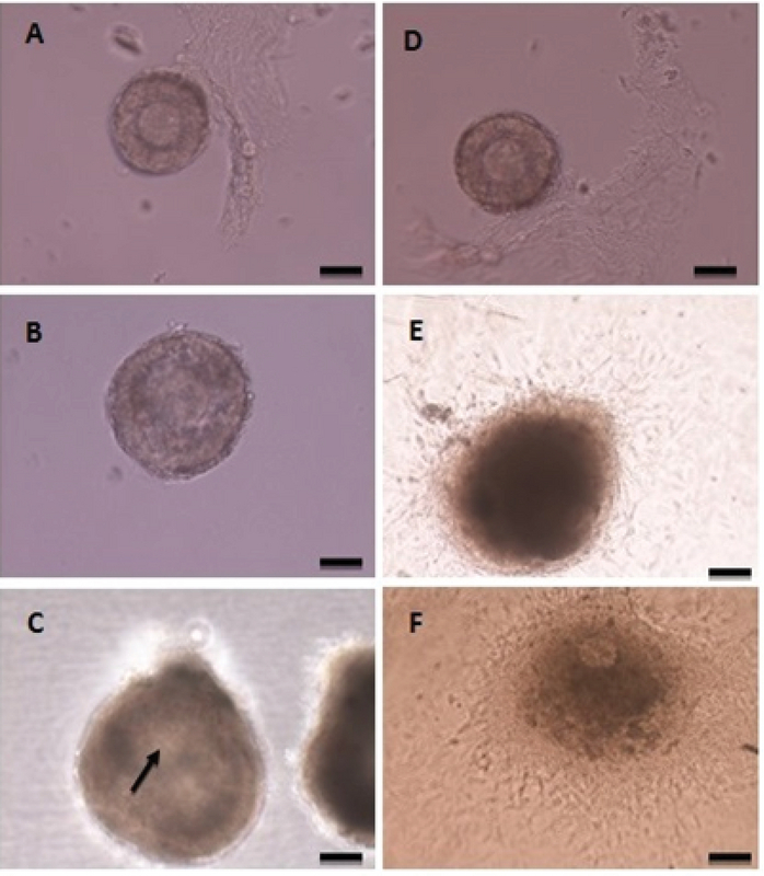
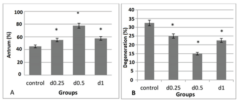
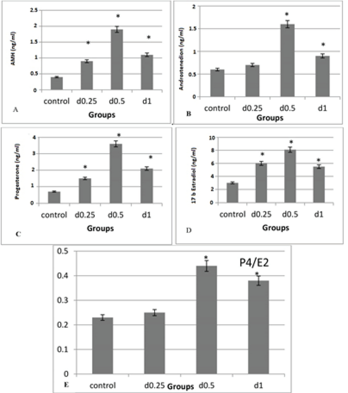
Immature female BALB/c mice (12-14 days) were sacrificed. The follicles were removed mechanically and transferred into α minimal essential medium with 5% fetal bovine serum. The preantral follicles were incubated with different concentrations of sodium alginate (0.25%, 0.5%, and 1%) and 2D medium for 12 days. The follicles were examined for antral formation following the 10th day and the diameter on days 6 and 12 . The levels of hormones (AMH, androstenedione, 17 -estradiol, and progesterone) and the expression of genes (, , , , and ) at the end of the 12 day.
Maximum follicle diameter and highest percentage of antrum formation were related to 0.5% concentration (p = 0.00). The levels of hormones in different doses of sodium alginate were increased significantly compared to the control group (p = 0.00). The highest and lowest levels of these hormones were related to 0.5% concentration and 2D medium, respectively. The highest level of genes expression was observed in 0.5% sodium alginate, which showed a significant increase compared to the control group (p = 0.00).
Proper concentration of alginate hydrogel increases follicle growth, causes follicle maturation, produces steroid hormones, and increases appropriate expression of steroidogenesis-related genes.
体外成熟方法的改进能够激活窦前卵泡生长,在生殖技术中成熟卵母细胞的产生方面发挥着关键作用。
评估不同浓度的海藻酸钠三维支架对小鼠窦前卵泡中激素和基因表达的影响。
处死未成熟雌性BALB/c小鼠(12 - 14日龄)。机械分离卵泡并转移至含5%胎牛血清的α - 最低必需培养基中。将窦前卵泡与不同浓度的海藻酸钠(0.25%、0.5%和1%)及二维培养基孵育12天。在第10天后检查卵泡的腔形成情况,并测量第6天和第12天的直径。在第12天结束时检测激素(抗苗勒管激素、雄烯二酮、17β - 雌二醇和孕酮)水平以及基因(相关基因名称未给出,此处保留原文)的表达。
最大卵泡直径和最高腔形成百分比与0.5%浓度相关(p = 0.00)。与对照组相比,不同剂量海藻酸钠组的激素水平显著升高(p = 0.00)。这些激素的最高和最低水平分别与0.5%浓度和二维培养基相关。在0.5%海藻酸钠组中观察到基因表达水平最高,与对照组相比显著增加(p = 0.00)。
适当浓度的海藻酸盐水凝胶可促进卵泡生长,导致卵泡成熟,产生甾体激素,并增加甾体生成相关基因的适当表达。