Universidade de São Paulo, Faculdade de Odontologia de Ribeirão Preto, Divisão de Ortodontia, Ribeirão Preto, SP, Brazil.
Universidade de São Paulo, Faculdade de Medicina de Ribeirão Preto, Hospital das Clínicas, Divisão de Fonoaudiologia, Ribeirão Preto, SP, Brazil.
Braz J Otorhinolaryngol. 2022 May-Jun;88(3):331-336. doi: 10.1016/j.bjorl.2020.06.008. Epub 2020 Jul 27.
The association between the intensity of obstructive sleep apnea and skeletal alterations in the face and hyoid bone is still scarcely addressed in the literature.
To evaluate whether the intensity of obstructive sleep apnea is associated with craniofacial alterations and the position of the hyoid bone in children with mixed dentition.
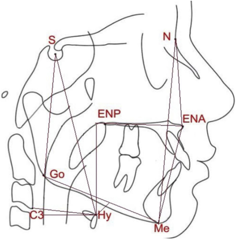
76 children aged 7 to 10 years old were examined by otorhinolaryngological evaluation, polysomnography, and orthodontic assessment, including cephalometry. The participants were divided in 3 groups: primary snoring, mild obstructive sleep apnea and moderate to severe obstructive sleep apnea. Cephalometric measures of the face and hyoid bone were assessed. These measures were compared among the different groups by unpaired Student's t test. Moreover, these measures were correlated with the patient's obstructive apnea and hypopnea index variable using Pearson's correlation test.
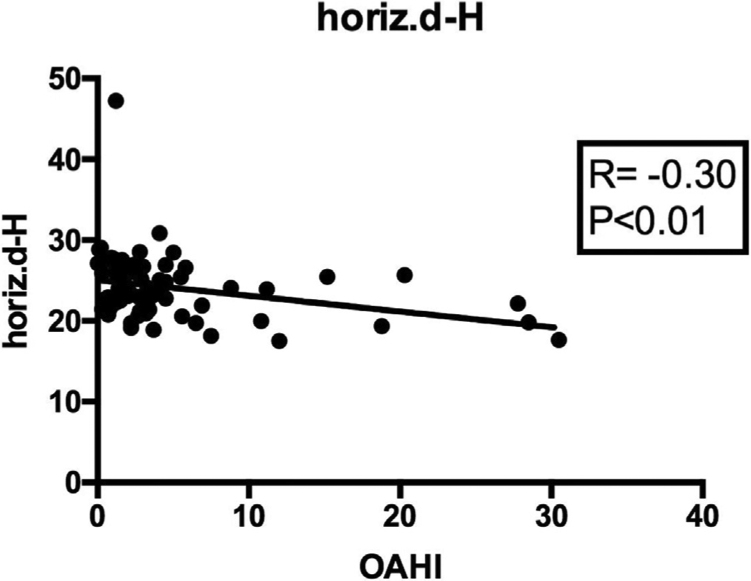
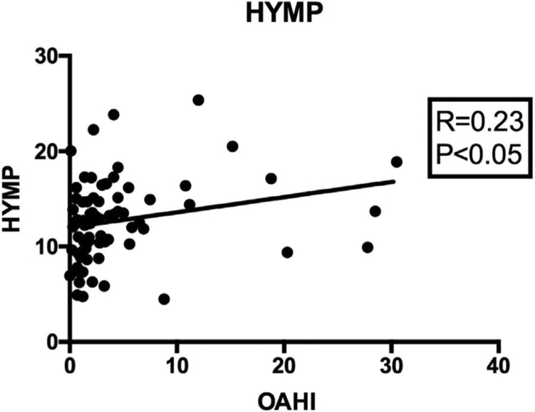
Of the 76 children, 14 belonged to group 1, with primary snoring; 46 to group 2, with mild obstructive sleep apnea; and 16 to group 3, with moderate-severe obstructive sleep apnea. There was no difference between the groups regarding the craniofacial variables. Children with obstructive sleep apnea showed a longer distance from the hyoid bone to the mandibular plane when compared to the primary snoring group (p<0.05). Between the two obstructive sleep apnea subgroups, patients with moderate or severe disease showed significantly shorter horizontal distance between the hyoid bone and the posterior pharyngeal wall (p<0.05), when compared to the groups with mild obstructive sleep apnea. We also observed a significant positive correlation between obstructive apnea and hypopnea index and the distance from the hyoid to the mandibular plane (p<0.05) as well as a significant negative association between obstructive apnea and hypopnea index and the horizontal distance from the hyoid to the posterior pharyngeal wall (p<0.01).
We did not observe any association between obstructive sleep apnea and linear lateral alterations of the face. In contrast, there is a direct association between obstructive sleep apnea severity and the inferior and posterior position of the hyoid bone in children aged 7 to 10 years old.
阻塞性睡眠呼吸暂停的严重程度与颌面和舌骨骨骼改变之间的关系在文献中仍鲜有报道。
评估阻塞性睡眠呼吸暂停的严重程度是否与混合牙列期儿童的颌面改变和舌骨位置有关。
对 76 名 7 至 10 岁的儿童进行耳鼻喉科评估、多导睡眠图和正畸评估,包括头影测量。将参与者分为三组:单纯性打鼾、轻度阻塞性睡眠呼吸暂停和中重度阻塞性睡眠呼吸暂停。评估面部和舌骨的头影测量值。通过独立样本 t 检验比较不同组之间的这些测量值。此外,还使用 Pearson 相关检验将这些测量值与患者的阻塞性呼吸暂停和低通气指数变量相关联。
76 名儿童中,14 名属于单纯性打鼾组(第 1 组),46 名属于轻度阻塞性睡眠呼吸暂停组(第 2 组),16 名属于中重度阻塞性睡眠呼吸暂停组(第 3 组)。三组间颅面变量无差异。与单纯性打鼾组相比,阻塞性睡眠呼吸暂停组的舌骨至下颌平面的距离更长(p<0.05)。在两个阻塞性睡眠呼吸暂停亚组之间,与轻度阻塞性睡眠呼吸暂停组相比,中重度疾病患者的舌骨与后咽壁之间的水平距离明显更短(p<0.05)。我们还观察到阻塞性呼吸暂停和低通气指数与舌骨至下颌平面的距离之间存在显著正相关(p<0.05),以及阻塞性呼吸暂停和低通气指数与舌骨至后咽壁的水平距离之间存在显著负相关(p<0.01)。
我们没有观察到阻塞性睡眠呼吸暂停与颌面线性侧方改变之间存在任何关联。相反,在 7 至 10 岁儿童中,阻塞性睡眠呼吸暂停的严重程度与舌骨的下后位置存在直接关联。