Su Wei, Tai Yang, Tang Shi-Hang, Ye Yan-Ting, Zhao Chong, Gao Jin-Hang, Tuo Bi-Guang, Tang Cheng-Wei
Department of Gastroenterology, West China Hospital, Sichuan University, Chengdu 610041, Sichuan Province, China.
Laboratory of Gastroenterology and Hepatology, West China Hospital, Sichuan University, Chengdu 610041, Sichuan Province, China.
World J Gastroenterol. 2020 Jul 28;26(28):4094-4107. doi: 10.3748/wjg.v26.i28.4094.
Endoplasmic reticulum (ER) stress is an important mechanism in the progression of chronic and acute liver diseases, especially in the progression and recovery of liver fibrosis. Excessive and long-term ER stress induces apoptosis. ER stress-induced apoptosis is considered to be an important pathway in the development of liver fibrosis. Cyclooxygenase-2 (COX-2) induction is also closely related to ER stress. In our previous studies, we showed that celecoxib, a COX-2 inhibitor, improves liver fibrosis and portal hypertension. However, the role and mechanism of celecoxib in alleviating liver fibrosis remain unclear.
To investigate whether celecoxib alleviates liver fibrosis by inhibiting hepatocyte apoptosis the ER stress response.
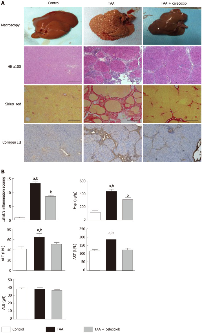
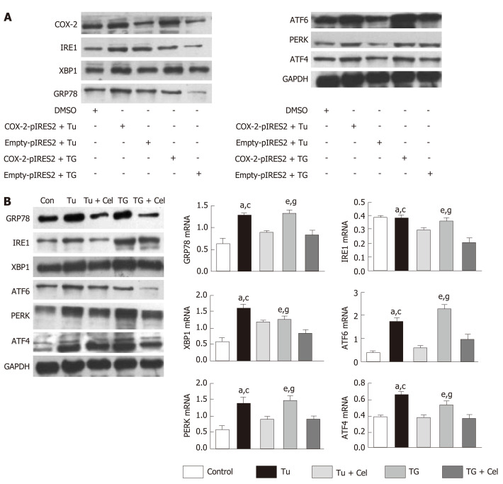
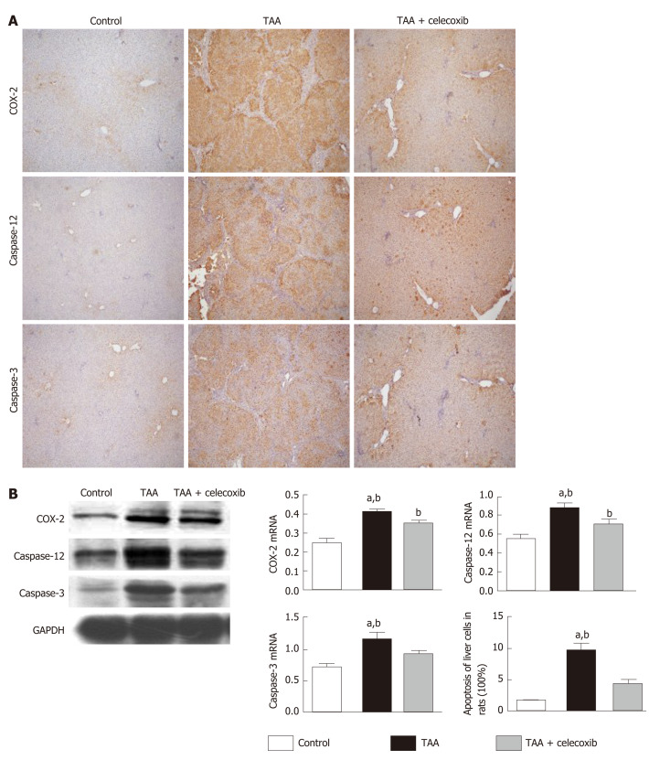
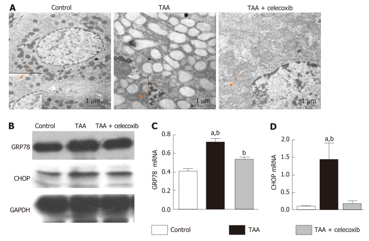
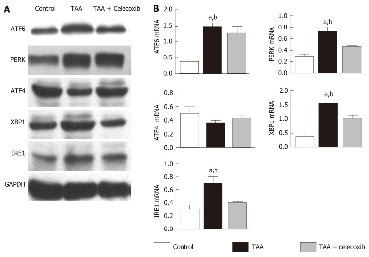
Cirrhosis was induced by intraperitoneal injections of thioacetamide (TAA) for 16 wk (injection dose is 200 mg/kg per 3 d for the first 8 wk and 100 mg /kg per 3 d after 8 wk). Thirty-six male Sprague-Dawley rats were randomly divided into three groups, namely, control group, TAA group, and TAA + celecoxib group. In the last 8 wk, TAA-induced cirrhotic rats received celecoxib (20 mg/kg/day) or the vehicle by gastric gavage. After 16 wk, the rats were sacrificed, and serum alanine aminotransferase (ALT), aspartate aminotransferase (AST), and albumin (ALB) were detected. The hepatic fibrosis areas were evaluated by Sirius red staining and the degree of fibrosis was assessed by measuring the level of hydroxyproline. ER stress levels were evaluated by detecting the marker proteins glucose-regulated protein 78 (GRP78), CCAAT/enhancer binding protein homologous protein (CHOP), PKR-like ER protein kinase (PERK), activating transcription factor 6 (ATF6), and inositol-requiring enzyme 1 alpha (IRE1α). Apoptosis levels were evaluated by detecting caspase-12 and caspase-3.
The serum ALT and AST levels in the liver were significantly reduced by celecoxib; however, the serum ALB had no significant changes. Celecoxib significantly reduced the degree of liver fibrosis and the levels of hydroxyproline (-38% and -25.7%, respectively, < 0.01). Celecoxib ameliorated ER stress by reducing the level of GRP78 compared to the TAA group ( < 0.05). Consistently, after celecoxib administration, the upregulation of TAA-induced hepatic apoptosis markers (caspase-12 and caspase-3) and CHOP were significantly inhibited. In addition, after celecoxib treatment, the expression of key molecules associated with ER stress (PERK, ATF6, and IRE1) was decreased ( < 0.05).
Therapeutic administration of celecoxib effectively reduces hepatic apoptosis in TAA-induced cirrhotic rats. The mechanism of action may be attributed to the suppression of CHOP expression, which subsequently inhibits ER stress.
内质网(ER)应激是慢性和急性肝病进展中的重要机制,尤其是在肝纤维化的进展和恢复过程中。过度和长期的内质网应激会诱导细胞凋亡。内质网应激诱导的细胞凋亡被认为是肝纤维化发展的重要途径。环氧合酶-2(COX-2)的诱导也与内质网应激密切相关。在我们之前的研究中,我们表明COX-2抑制剂塞来昔布可改善肝纤维化和门静脉高压。然而,塞来昔布在减轻肝纤维化中的作用和机制仍不清楚。
研究塞来昔布是否通过抑制肝细胞凋亡的内质网应激反应来减轻肝纤维化。
通过腹腔注射硫代乙酰胺(TAA)16周诱导肝硬化(注射剂量为前8周每3天200mg/kg,8周后每3天100mg/kg)。将36只雄性Sprague-Dawley大鼠随机分为三组,即对照组、TAA组和TAA+塞来昔布组。在最后8周,TAA诱导的肝硬化大鼠通过灌胃给予塞来昔布(20mg/kg/天)或溶剂。16周后,处死大鼠,检测血清丙氨酸氨基转移酶(ALT)、天冬氨酸氨基转移酶(AST)和白蛋白(ALB)。通过天狼星红染色评估肝纤维化面积,并通过测量羟脯氨酸水平评估纤维化程度。通过检测标记蛋白葡萄糖调节蛋白78(GRP78)、CCAAT/增强子结合蛋白同源蛋白(CHOP)、PKR样内质网蛋白激酶(PERK)、活化转录因子6(ATF6)和肌醇需求酶1α(IRE1α)评估内质网应激水平。通过检测半胱天冬酶-12和半胱天冬酶-3评估细胞凋亡水平。
塞来昔布显著降低了肝脏中血清ALT和AST水平;然而,血清ALB没有显著变化。塞来昔布显著降低了肝纤维化程度和羟脯氨酸水平(分别降低了38%和25.7%,P<0.01)。与TAA组相比,塞来昔布通过降低GRP78水平改善了内质网应激(P<0.05)。同样,给予塞来昔布后,TAA诱导的肝细胞凋亡标志物(半胱天冬酶-12和半胱天冬酶-3)和CHOP的上调被显著抑制。此外,塞来昔布治疗后,与内质网应激相关的关键分子(PERK、ATF6和IRE1)的表达降低(P<0.05)。
塞来昔布治疗可有效降低TAA诱导的肝硬化大鼠的肝细胞凋亡。其作用机制可能归因于CHOP表达的抑制,随后抑制了内质网应激。