Wang Xin-Xin, Shao Chen, Huang Xiao-Jie, Sun Lin, Meng Ling-Jia, Liu Hui, Zhang Shi-Jie, Li Hong-Jun, Lv Fu-Dong
Department of Pathology, Beijing Youan Hospital, Capital Medical University, Beijing, China.
Department of Infectious Disease, Beijing Youan Hospital, Capital Medical University, Beijing, China.
J Clin Pathol. 2021 Aug;74(8):522-527. doi: 10.1136/jclinpath-2020-206623. Epub 2020 Aug 26.
The global outbreak of COVID-19 has resulted in an increased mortality. However, whether severe acute respiratory syndrome coronavirus 2 (SARS-CoV-2) can affect multiple organs is still unclear. In this study, postmortem percutaneous biopsies of multiple organs from deceased patients were performed to understand the histopathological changes caused by COVID-19.
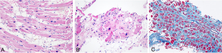
Biopsy specimens of pulmonary, cardiac, hepatic and lymphoid tissues were obtained from three patients, who died due to COVID-19 pneumonia. H&E stain, Masson trichrome stain, immunohistochemistry stain and in-situ hybridisation were used.
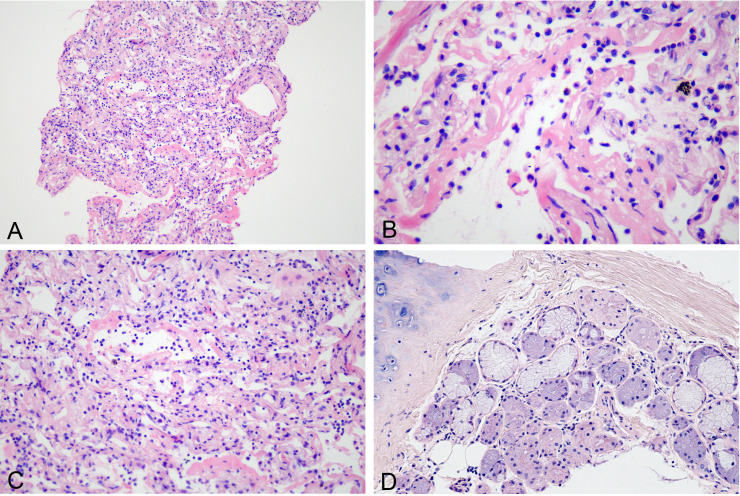
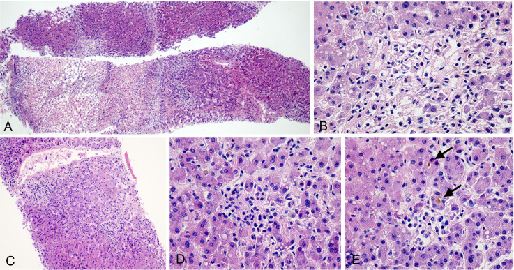
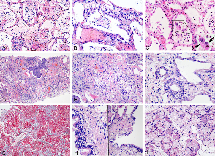
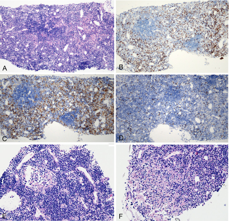
Pulmonary damages caused by SARS-CoV-2 infection was diffuse alveolar damage (DAD). In the early phase, the histological findings were mainly those of exudative features of DAD. The later phase was characterised by organisation of DAD combined with bacterial pneumonia. No serious damage was found in the bronchiolar epithelium and submucosal glands. The hepatic tissue revealed features of ischaemic necrosis, but findings suggestive of mild lobular hepatitis were also observed. The lymphoid tissue revealed features of non-specific acute lymphadenitis. The cardiac tissue revealed changes of underlying disease. SARS-CoV-2 RNAs were not detected in hepatocytes, cholangiocytes and lymphocytes of lymph nodes.
COVID-19 predominantly involves the pulmonary tissue, causes DAD and aggravates the cardiovascular disease. However, other extrapulmonary tissues did not reveal any virus-specific findings, but were affected by multiple factors. The findings in this report caution the pathologists that they should not mistakenly attribute all the histological features to CoV infection. Moreover, the clinicians should pay attention to the potentially injurious and correctable causes.
新型冠状病毒肺炎(COVID-19)的全球大流行导致死亡率上升。然而,严重急性呼吸综合征冠状病毒2(SARS-CoV-2)是否会影响多个器官仍不清楚。在本研究中,对死亡患者的多个器官进行了尸检经皮活检,以了解COVID-19引起的组织病理学变化。
从3例因COVID-19肺炎死亡的患者获取肺、心脏、肝脏和淋巴组织的活检标本。采用苏木精-伊红(H&E)染色、Masson三色染色、免疫组织化学染色和原位杂交。
SARS-CoV-2感染引起的肺部损伤为弥漫性肺泡损伤(DAD)。早期,组织学表现主要为DAD的渗出性特征。后期以DAD的机化合并细菌性肺炎为特征。细支气管上皮和黏膜下腺未发现严重损伤。肝组织表现为缺血性坏死特征,但也观察到轻度小叶性肝炎的表现。淋巴组织表现为非特异性急性淋巴结炎特征。心脏组织表现为基础疾病的改变。在肝细胞、胆管细胞和淋巴结淋巴细胞中未检测到SARS-CoV-2 RNA。
COVID-19主要累及肺组织,引起DAD并加重心血管疾病。然而,其他肺外组织未发现任何病毒特异性表现,但受多种因素影响。本报告中的发现提醒病理学家,不应将所有组织学特征错误地归因于冠状病毒感染。此外,临床医生应注意潜在的有害且可纠正的病因。