Ullah Anwar, Masood Rehana
Department of Biosciences, COMSATS University Islamabad, Islamabad, Pakistan.
Department of Biochemistry, Shaheed Benazir Bhutto Women University Peshawar, Peshawar, Pakistan.
Front Mol Biosci. 2020 Aug 5;7:175. doi: 10.3389/fmolb.2020.00175. eCollection 2020.
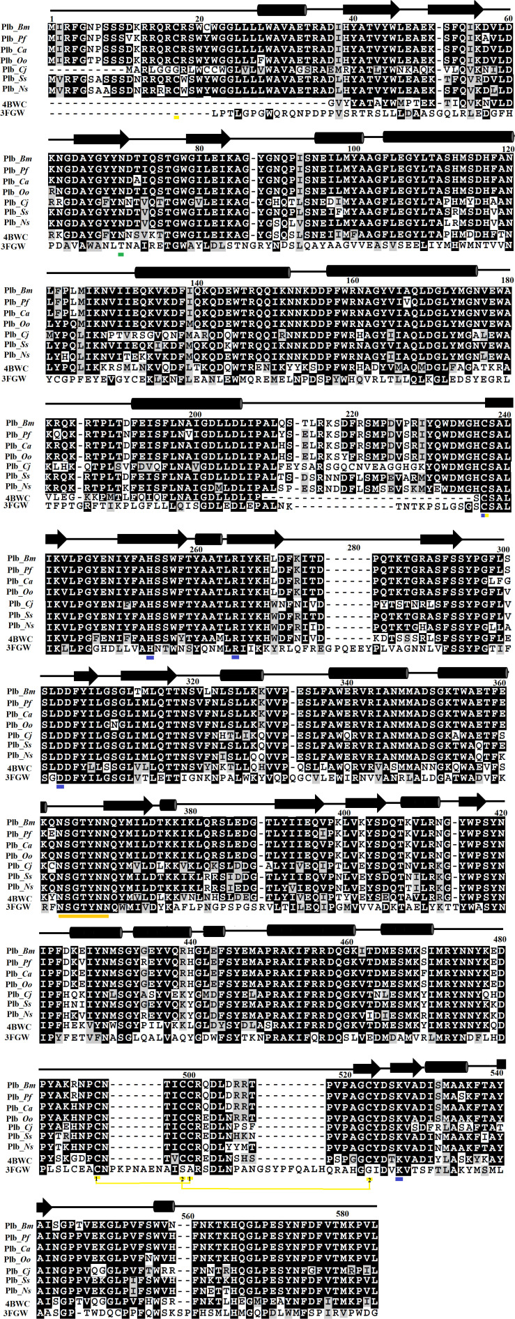
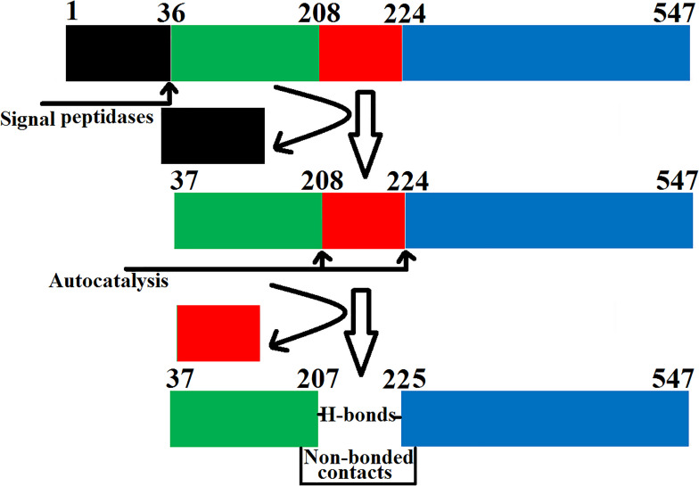
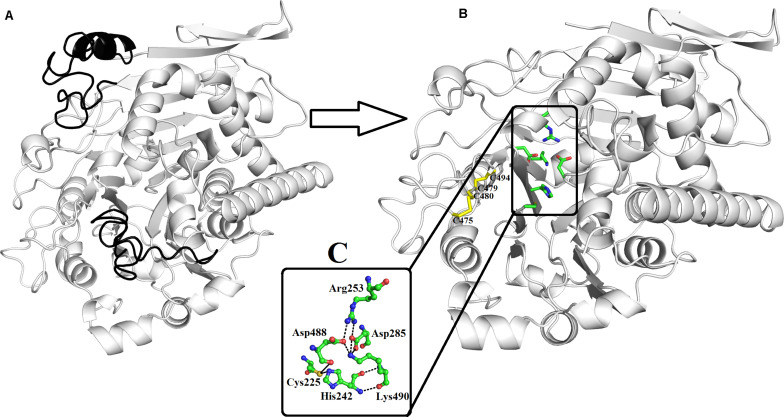
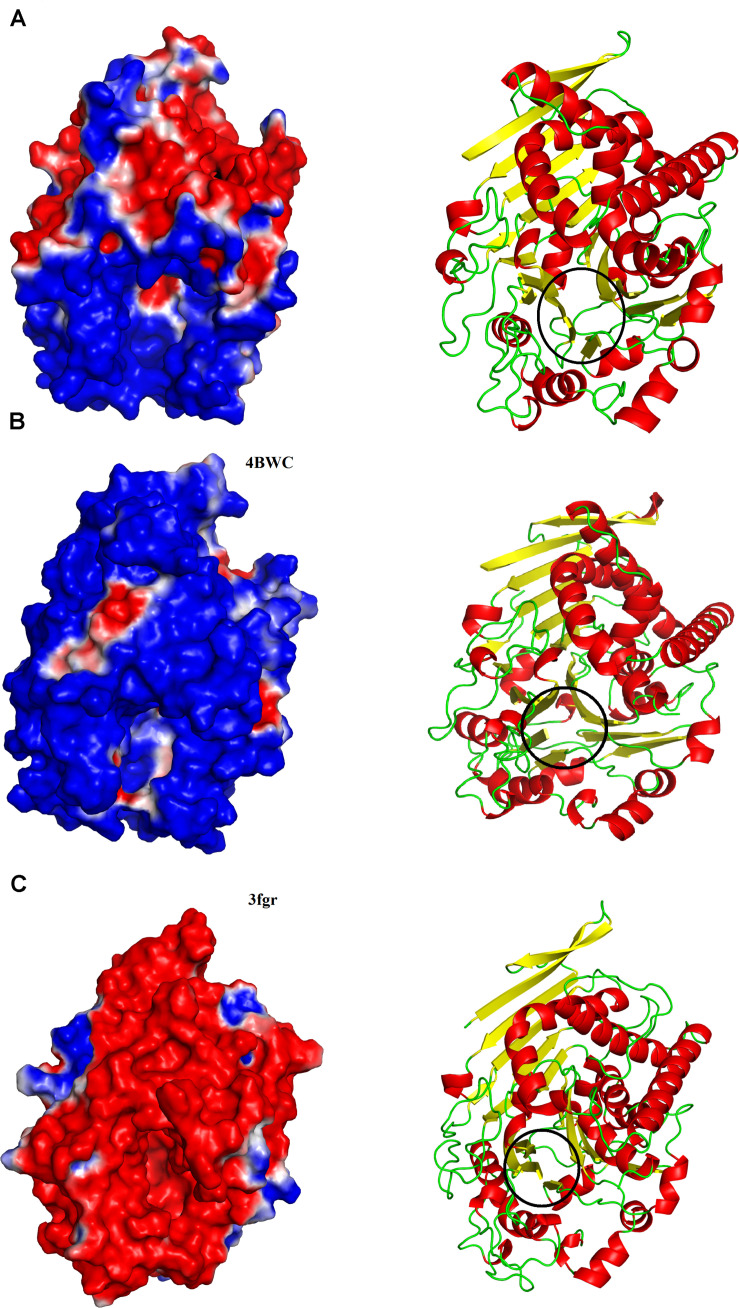
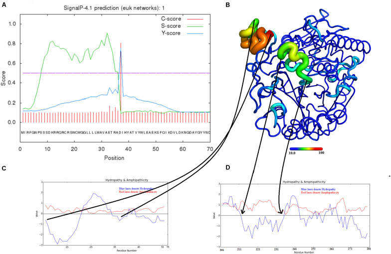
Snake venom phospholipases B (SVPLBs) are the least studied enzymes. They constitute about 1% of crude venoms, however, in other snake venoms, it is present in less than 1%. These enzymes are considered the most potent hemolytic agent in the venom. Currently, no structural information is available about these enzymes from snake venom. To better understand its three-dimensional structure and mechanisms of envenomation, the current work describes the first model-based structure report of this enzyme from venom named as phospholipase B (PLB_). The structure model of PLB_ was generated using model building software like I-TESSER, MODELLER 9v19, and Swiss-Model. The build PLB_ model was validated using validation tools (PROCHECK, ERRAT, and Verif3D). The analysis of the PLB_ modeled structure indicates that it contains 491 amino acid residues that form a well-defined four-layer αββα sandwich core and has a typical fold of the N-terminal nucleophile aminohydrolase (Ntn-hydrolase). The overall structure of PLB_ contains 18 β-strands and 17 α-helices with many connecting loops. The structure divides into two chains (A and B) after maturation. The A chain is smaller and contains 207 amino acid residues, whereas the B chain is larger and contains 266 amino acid residues. The sequence and structural comparison among homologous snake venom, bacterial, and mammals PLBs indicate that differences in the length and sequence composition may confer variable substrate specificity to these enzymes. Moreover, the surface charge distribution, average volume, and depth of the active site cavity also vary in these enzymes. The present work will provide more information about the structure-function relationship and mechanism of action of these enzymes in snakebite envenomation.
蛇毒磷脂酶B(SVPLBs)是研究最少的酶。它们约占粗毒液的1%,然而,在其他蛇毒中,其含量不到1%。这些酶被认为是毒液中最有效的溶血剂。目前,尚无关于这些蛇毒酶的结构信息。为了更好地了解其三维结构和中毒机制,目前的工作描述了这种来自毒液的名为磷脂酶B(PLB_)的酶的首个基于模型的结构报告。PLB_的结构模型是使用I-TESSER、MODELLER 9v19和Swiss-Model等模型构建软件生成的。构建的PLB_模型使用验证工具(PROCHECK、ERRAT和Verif3D)进行了验证。对PLB_建模结构的分析表明,它包含491个氨基酸残基,形成了一个定义明确的四层αββα夹心核心,具有典型的N-末端亲核氨基水解酶(Ntn-水解酶)折叠。PLB_的整体结构包含18条β链和17个α螺旋,有许多连接环。成熟后,该结构分为两条链(A链和B链)。A链较小,包含207个氨基酸残基,而B链较大,包含266个氨基酸残基。同源蛇毒、细菌和哺乳动物PLBs之间的序列和结构比较表明,长度和序列组成的差异可能赋予这些酶不同的底物特异性。此外,这些酶的表面电荷分布、平均体积和活性位点腔的深度也有所不同。目前的工作将提供更多关于这些酶在蛇咬伤中毒中的结构-功能关系和作用机制的信息。