Laboratory of Molecular Imaging and Nanomedicine, National Institute of Biomedical Imaging and Bioengineering, National Institutes of Health, Bethesda, MD 20892, USA.
Laboratory of Molecular Imaging and Nanomedicine, National Institute of Biomedical Imaging and Bioengineering, National Institutes of Health, Bethesda, MD 20892, USA.
EBioMedicine. 2020 Sep;59:102958. doi: 10.1016/j.ebiom.2020.102958. Epub 2020 Aug 25.
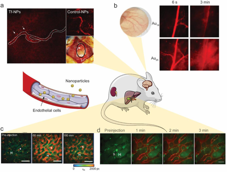
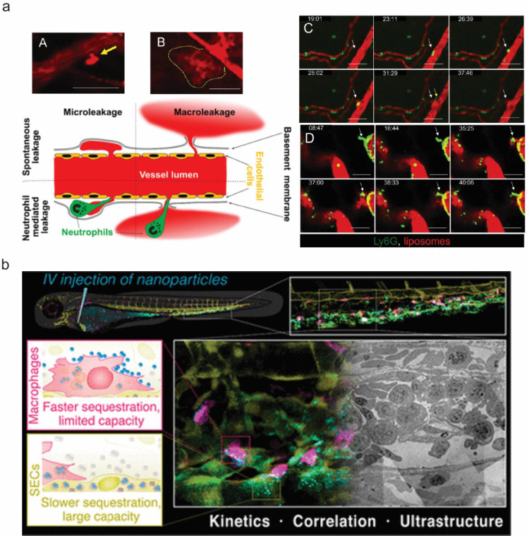
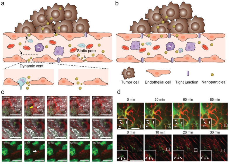
Nanomedicine has proven promising in preclinical studies. However, only few formulations have been successfully translated to clinical use. A thorough understanding of how nanoparticles interact with cells in vivo is essential to accelerate the clinical translation of nanomedicine. Intravital imaging is a crucial tool to reveal the mechanisms of nanoparticle transport in vivo, allowing for the development of new strategies for nanomaterial design. Here, we first review the most recent progress in using intravital imaging to answer fundamental questions about nanoparticle delivery in vivo. We then elaborate on how nanoparticles interact with different cell types and how such interactions determine the fate of nanoparticles in vivo. Lastly, we discuss ways in which the use of intravital imaging can be expanded in the future to facilitate the clinical translation of nanomedicine.
纳米医学在临床前研究中已被证明具有广阔的前景。然而,只有少数制剂成功转化为临床应用。深入了解纳米颗粒如何与体内细胞相互作用对于加速纳米医学的临床转化至关重要。活体成像技术是揭示纳米颗粒在体内运输机制的重要工具,有助于为纳米材料设计开发新策略。在这里,我们首先回顾了利用活体成像来回答关于纳米颗粒体内递释的基本问题的最新进展。然后,我们详细阐述了纳米颗粒如何与不同类型的细胞相互作用,以及这种相互作用如何决定纳米颗粒在体内的命运。最后,我们讨论了未来如何扩大活体成像的应用范围,以促进纳米医学的临床转化。