• 文献检索
  • 文档翻译
  • 深度研究
  • 学术资讯
  • Suppr Zotero 插件Zotero 插件
  • 邀请有礼
  • 套餐&价格
  • 历史记录
应用&插件
Suppr Zotero 插件Zotero 插件浏览器插件Mac 客户端Windows 客户端微信小程序
定价
高级版会员购买积分包购买API积分包
服务
文献检索文档翻译深度研究API 文档MCP 服务
关于我们
关于 Suppr公司介绍联系我们用户协议隐私条款
关注我们

Suppr 超能文献

核心技术专利:CN118964589B侵权必究
粤ICP备2023148730 号-1Suppr @ 2026

文献检索

告别复杂PubMed语法,用中文像聊天一样搜索,搜遍4000万医学文献。AI智能推荐,让科研检索更轻松。

立即免费搜索

文件翻译

保留排版,准确专业,支持PDF/Word/PPT等文件格式,支持 12+语言互译。

免费翻译文档

深度研究

AI帮你快速写综述,25分钟生成高质量综述,智能提取关键信息,辅助科研写作。

立即免费体验

使用不同人血清扩增人角膜缘上皮干细胞/祖细胞:多变量统计分析。

Expansion of Human Limbal Epithelial Stem/Progenitor Cells Using Different Human Sera: A Multivariate Statistical Analysis.

机构信息

Department of Cell Biology and Histology, School of Medicine and Nursing, Biocruces Bizkaia Health Research Institute, University of the Basque Country UPV/EHU, 48940 Leioa, Bizkaia, Spain.

Cornea Division, Stein Eye Institute, University of California, Los Angeles, CA 90095, USA.

出版信息

Int J Mol Sci. 2020 Aug 25;21(17):6132. doi: 10.3390/ijms21176132.

DOI:10.3390/ijms21176132
PMID:32854428
原文链接:https://pmc.ncbi.nlm.nih.gov/articles/PMC7503296/
Abstract

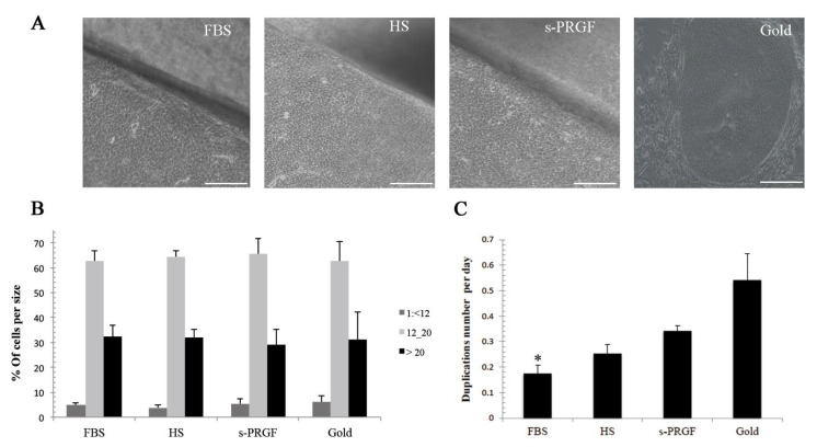
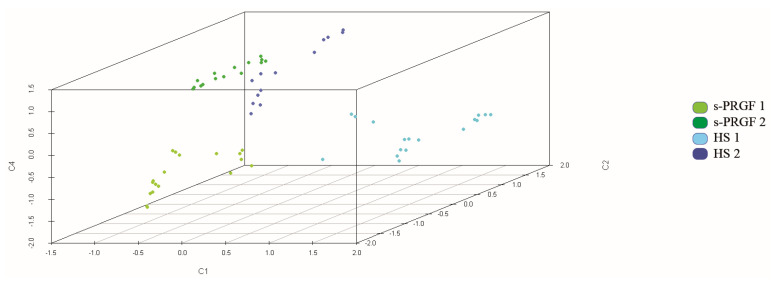
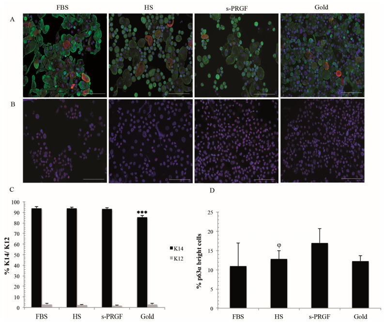
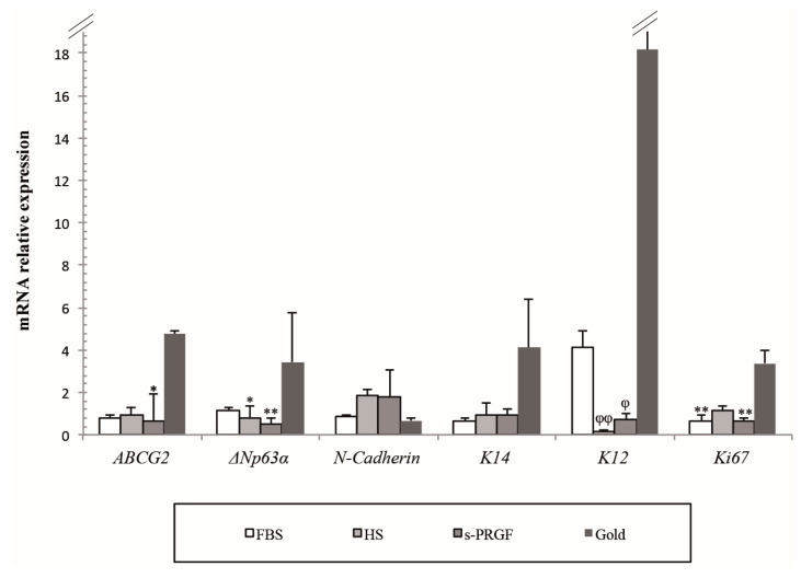
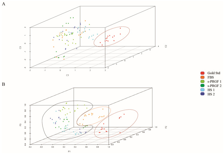
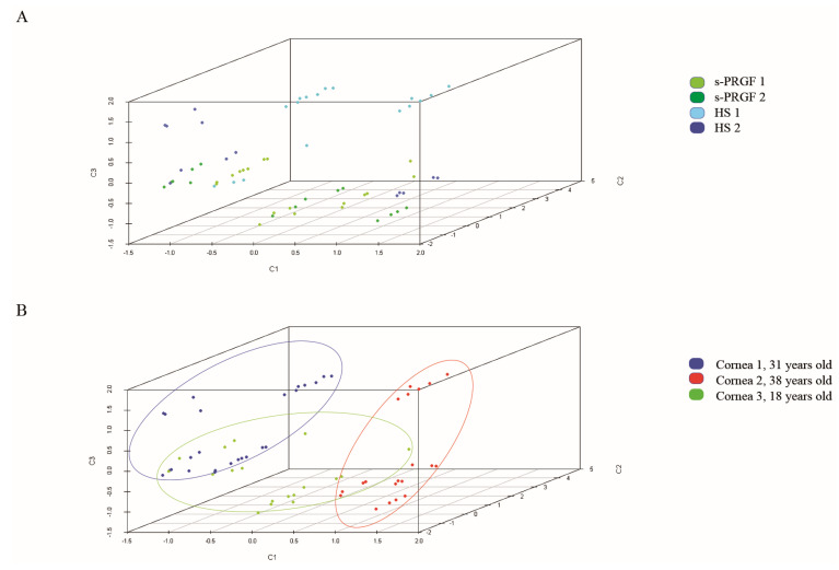
Transplantation of human cultured limbal epithelial stem/progenitor cells (LESCs) has demonstrated to restore the integrity and functionality of the corneal surface in about 76% of patients with limbal stem cell deficiency. However, there are different protocols for the expansion of LESCs, and many of them use xenogeneic products, being a risk for the patients' health. We compared the culture of limbal explants on the denuded amniotic membrane in the culture medium-supplemental hormone epithelial medium (SHEM)-supplemented with FBS or two differently produced human sera. Cell morphology, cell size, cell growth rate, and the expression level of differentiation and putative stem cell markers were examined. Several bioactive molecules were quantified in the human sera. In a novel approach, we performed a multivariate statistical analysis of data to investigate the culture factors, such as differently expressed molecules of human sera that specifically influence the cell phenotype. Our results showed that limbal cells cultured with human sera grew faster and contained similar amounts of small-sized cells, higher expression of the protein p63α, and lower of cytokeratin K12 than FBS cultures, thus, maintaining the stem/progenitor phenotype of LESCs. Furthermore, the multivariate analysis provided much data to better understand the obtaining of different cell phenotypes as a consequence of the use of different culture methodologies or different culture components.

摘要

人培养的角膜缘上皮干细胞/祖细胞(LESCs)移植已证明可恢复约 76%的角膜缘干细胞缺乏症患者的角膜表面完整性和功能。然而,LESCs 的扩增有不同的方案,其中许多方案使用异种产品,这对患者的健康构成风险。我们比较了在补充有 FBS 的基础激素上皮培养基(SHEM)中的裸羊膜上培养角膜缘组织片,或在两种不同来源的人血清中的培养。检测了细胞形态、细胞大小、细胞生长速度以及分化和潜在干细胞标志物的表达水平。在人血清中定量了几种生物活性分子。在一种新方法中,我们对数据进行了多变量统计分析,以研究培养因素,如人血清中差异表达的分子,这些分子特异性地影响细胞表型。我们的结果表明,用人血清培养的角膜缘细胞生长更快,含有更多数量的小细胞,p63α 蛋白表达更高,细胞角蛋白 K12 表达更低,因此保持了 LESCs 的干细胞/祖细胞表型。此外,多元分析提供了更多的数据,有助于更好地理解由于使用不同的培养方法学或不同的培养成分而获得不同的细胞表型。

https://cdn.ncbi.nlm.nih.gov/pmc/blobs/0c5a/7503296/b1e805d2894e/ijms-21-06132-g006.jpg
https://cdn.ncbi.nlm.nih.gov/pmc/blobs/0c5a/7503296/d028d0c087fc/ijms-21-06132-g001.jpg
https://cdn.ncbi.nlm.nih.gov/pmc/blobs/0c5a/7503296/daca806d5ee9/ijms-21-06132-g002.jpg
https://cdn.ncbi.nlm.nih.gov/pmc/blobs/0c5a/7503296/183b7bfcbbcf/ijms-21-06132-g003.jpg
https://cdn.ncbi.nlm.nih.gov/pmc/blobs/0c5a/7503296/09d7987810bd/ijms-21-06132-g004.jpg
https://cdn.ncbi.nlm.nih.gov/pmc/blobs/0c5a/7503296/d4ba33f800d5/ijms-21-06132-g005.jpg
https://cdn.ncbi.nlm.nih.gov/pmc/blobs/0c5a/7503296/b1e805d2894e/ijms-21-06132-g006.jpg

相似文献

1
Expansion of Human Limbal Epithelial Stem/Progenitor Cells Using Different Human Sera: A Multivariate Statistical Analysis.使用不同人血清扩增人角膜缘上皮干细胞/祖细胞:多变量统计分析。
Int J Mol Sci. 2020 Aug 25;21(17):6132. doi: 10.3390/ijms21176132.
2
Comparison of culture media for ex vivo cultivation of limbal epithelial progenitor cells.用于角膜缘上皮祖细胞体外培养的培养基比较
Mol Vis. 2013;19:69-77. Epub 2013 Jan 17.
3
Ex vivo expanded autologous limbal epithelial cells on amniotic membrane using a culture medium with human serum as single supplement.使用含有人血清作为唯一补充物的培养基,在羊膜上进行体外扩增自体角膜缘上皮细胞。
Exp Eye Res. 2012 Apr;97(1):1-9. doi: 10.1016/j.exer.2012.01.013. Epub 2012 Feb 7.
4
Human corneal epithelial subpopulations: oxygen dependent ex vivo expansion and transcriptional profiling.人眼角膜上皮亚群:依赖氧的体外扩增和转录谱分析。
Acta Ophthalmol. 2013 Jun;91 Thesis 4:1-34. doi: 10.1111/aos.12157.
5
Characterisation of corneas following different time and storage methods for their use as a source of stem-like limbal epithelial cells.不同时间和储存方法对角膜特性的影响,及其作为干细胞样角膜缘上皮细胞来源的应用。
Exp Eye Res. 2021 Oct;211:108720. doi: 10.1016/j.exer.2021.108720. Epub 2021 Aug 11.
6
Functional reconstruction of rabbit corneal epithelium by human limbal cells cultured on amniotic membrane.羊膜上培养的人角膜缘细胞对兔角膜上皮的功能重建
Mol Vis. 2003 Dec 8;9:635-43.
7
A simple and efficient method for cultivation of limbal explant stem cells with clinically safe potential.一种简单高效的培养角膜缘外植体干细胞的方法,具有临床安全潜力。
Cytotherapy. 2019 Jan;21(1):83-95. doi: 10.1016/j.jcyt.2018.11.005. Epub 2018 Nov 30.
8
Long-Term Cultures of Human Cornea Limbal Explants Form 3D Structures Ex Vivo - Implications for Tissue Engineering and Clinical Applications.人角膜缘外植体的长期培养可在体外形成三维结构——对组织工程和临床应用的启示
PLoS One. 2015 Nov 18;10(11):e0143053. doi: 10.1371/journal.pone.0143053. eCollection 2015.
9
Comparison of stem cell properties in cell populations isolated from human central and limbal corneal epithelium.比较从人中央和角膜缘角膜上皮分离的细胞群体中的干细胞特性。
Cornea. 2011 Oct;30(10):1155-62. doi: 10.1097/ICO.0b013e318213796b.
10
Xenofree generation of limbal stem cells for ocular surface advanced cell therapy.用于眼表高级细胞治疗的无饲养层的角膜缘干细胞的体外生成。
Stem Cell Res Ther. 2019 Dec 4;10(1):374. doi: 10.1186/s13287-019-1501-9.

引用本文的文献

1
Progress in Transdifferentiation of Autologous Alternative Cell Sources into Corneal Epithelial Cells.自体替代细胞来源向角膜上皮细胞转分化的研究进展
Stem Cell Rev Rep. 2025 Jan;21(1):226-235. doi: 10.1007/s12015-024-10808-1. Epub 2024 Oct 31.
2
Ocular Surface Regeneration by Limbal Stem Cells Therapies: State of the Art, Challenges, and Perspectives.通过角膜缘干细胞疗法实现眼表面再生:现状、挑战与展望。
Stem Cells Transl Med. 2023 Nov 3;12(11):714-719. doi: 10.1093/stcltm/szad058.
3
Concise Review: Bioengineering of Limbal Stem Cell Niche.

本文引用的文献

1
The limbus: Structure and function.角膜缘:结构与功能。
Exp Eye Res. 2020 Aug;197:108074. doi: 10.1016/j.exer.2020.108074. Epub 2020 Jun 2.
2
Xenofree generation of limbal stem cells for ocular surface advanced cell therapy.用于眼表高级细胞治疗的无饲养层的角膜缘干细胞的体外生成。
Stem Cell Res Ther. 2019 Dec 4;10(1):374. doi: 10.1186/s13287-019-1501-9.
3
Short- and Long-Term Results of Xenogeneic-Free Cultivated Autologous and Allogeneic Limbal Epithelial Stem Cell Transplantations.同种异体无饲养培养自体和同种异体角膜缘上皮干细胞移植的短期和长期结果。
简要综述:角膜缘干细胞微环境的生物工程
Bioengineering (Basel). 2023 Jan 12;10(1):111. doi: 10.3390/bioengineering10010111.
4
Therapeutic Strategies for Restoring Perturbed Corneal Epithelial Homeostasis in Limbal Stem Cell Deficiency: Current Trends and Future Directions.治疗策略:恢复角膜缘干细胞缺陷中失调的角膜上皮稳态:当前趋势和未来方向。
Cells. 2022 Oct 16;11(20):3247. doi: 10.3390/cells11203247.
5
Platelet-Rich Plasma as an Alternative to Xenogeneic Sera in Cell-Based Therapies: A Need for Standardization.富血小板血浆作为细胞疗法中异种血清的替代品:标准化的必要性。
Int J Mol Sci. 2022 Jun 11;23(12):6552. doi: 10.3390/ijms23126552.
Cornea. 2019 Dec;38(12):1543-1549. doi: 10.1097/ICO.0000000000002153.
4
Fibronectin regulates the self-renewal of rabbit limbal epithelial stem cells by stimulating the Wnt11/Fzd7/ROCK non-canonical Wnt pathway.纤连蛋白通过刺激 Wnt11/Fzd7/ROCK 非经典 Wnt 通路调节兔角膜缘上皮干细胞的自我更新。
Exp Eye Res. 2019 Aug;185:107681. doi: 10.1016/j.exer.2019.05.021. Epub 2019 May 28.
5
Hyaluronic Acid Combined with Serum Rich in Growth Factors in Corneal Epithelial Defects.透明质酸联合富含生长因子的血清治疗角膜上皮缺损。
Int J Mol Sci. 2019 Apr 3;20(7):1655. doi: 10.3390/ijms20071655.
6
Substance P inhibits high urea-induced apoptosis through the AKT/GSK-3β pathway in human corneal epithelial cells.P物质通过AKT/GSK-3β信号通路抑制高尿素诱导的人角膜上皮细胞凋亡。
J Cell Biochem. 2019 Jul;120(7):11342-11349. doi: 10.1002/jcb.28410. Epub 2019 Feb 6.
7
The Role of Nerve Growth Factor in Maintaining Proliferative Capacity, Colony-Forming Efficiency, and the Limbal Stem Cell Phenotype.神经生长因子在维持增殖能力、集落形成效率和角膜缘干细胞表型中的作用。
Stem Cells. 2019 Jan;37(1):139-149. doi: 10.1002/stem.2921.
8
Comparative study of substrate free and amniotic membrane scaffolds for cultivation of limbal epithelial sheet.无载体和羊膜支架培养角膜缘上皮细胞片的对比研究。
Sci Rep. 2018 Oct 2;8(1):14628. doi: 10.1038/s41598-018-32914-0.
9
Characterization and comparison of human limbal explant cultures grown under defined and xeno-free conditions.在定义和无动物来源条件下培养的人角膜缘外植体的特征描述和比较。
Exp Eye Res. 2018 Nov;176:20-28. doi: 10.1016/j.exer.2018.06.019. Epub 2018 Jun 19.
10
Phase II Randomized, Double-Masked, Vehicle-Controlled Trial of Recombinant Human Nerve Growth Factor for Neurotrophic Keratitis.人神经生长因子治疗神经营养性角膜病变的随机、双盲、载体对照 II 期临床试验
Ophthalmology. 2018 Sep;125(9):1332-1343. doi: 10.1016/j.ophtha.2018.02.022. Epub 2018 Apr 10.