Jo Uiree, Song Joon Seon, Choi Seung-Ho, Nam Soon Yuhl, Kim Sang Yoon, Cho Kyung-Ja
Department of Pathology, Asan Medical Center, University of Ulsan College Medicine, Seoul, Korea.
Department of Otorhinolaryngology-Head and Neck Surgery, Asan Medical Center, University of Ulsan College Medicine, Seoul, Korea.
J Pathol Transl Med. 2020 Nov;54(6):489-496. doi: 10.4132/jptm.2020.07.19. Epub 2020 Aug 31.
Primary squamous cell carcinoma (SCC) of the salivary gland is a rare disease, and distinguishing primary SCC from metastatic SCC is difficult. This study investigated the histological and immunohistochemical differences between primary and metastatic salivary gland SCC to improve the accuracy of diagnosis and to explore the pathogenesis of this disease.
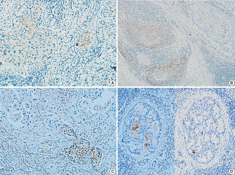
Data of 16 patients who underwent surgery for SCC of salivary glands between 2000 and 2018 at Asan Medical Center were retrieved. Eight patients had a history of SCC at other sites, and eight patients had only salivary gland SCC. Immunostaining for p16, p53, androgen receptor (AR), gross cystic disease fluid protein 15 (GCDFP-15), and c-erbB2, as well as mucicarmine staining, were compared between the two groups.
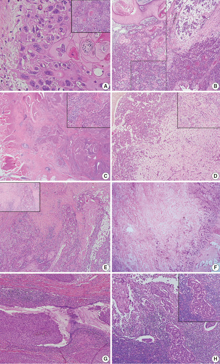
Most tumors were located in the center of the salivary glands with extraparenchymal extension. The histology of primary SCC of the salivary gland was consistent with moderately differentiated SCC with extensive desmoplastic reaction and peritumoral inflammation. Involvement of the salivary gland ducts and transition into the ductal epithelium were observed in two cases. Metastatic SCC resembled the primary tumor histologically and was associated with central necrosis. Both groups exhibited negative mucin staining. Two, one, and one primary SCC case exhibited AR, GCDFP-15, and c-erbB2 positivity, respectively.
A subset of primary SCCs originated in salivary ducts or was related to salivary duct carcinoma. Distinguishing primary from metastatic SCC of the salivary gland is difficult using histologic features and immunoprofiles. A comprehensive review of the medical history is essential.
涎腺原发性鳞状细胞癌(SCC)是一种罕见疾病,鉴别原发性SCC与转移性SCC具有一定难度。本研究调查原发性和转移性涎腺SCC之间的组织学和免疫组化差异,以提高诊断准确性并探索该疾病的发病机制。
检索2000年至2018年在峨山医学中心接受涎腺SCC手术的16例患者的数据。8例患者有其他部位SCC病史,8例患者仅有涎腺SCC。比较两组患者p16、p53、雄激素受体(AR)、巨大囊肿病液体蛋白15(GCDFP-15)和c-erbB2的免疫染色以及黏液卡红染色情况。
大多数肿瘤位于涎腺中央并向腺体外延伸。涎腺原发性SCC的组织学表现与中度分化的SCC一致,伴有广泛的促纤维组织增生反应和瘤周炎症。2例观察到涎腺导管受累并向导管上皮转变。转移性SCC在组织学上与原发性肿瘤相似,并伴有中央坏死。两组均表现为黏液染色阴性。原发性SCC分别有2例、1例和1例表现为AR、GCDFP-15和c-erbB2阳性。
一部分原发性SCC起源于涎腺导管或与涎腺导管癌相关。利用组织学特征和免疫表型鉴别涎腺原发性和转移性SCC具有一定难度。全面回顾病史至关重要。