Department of Biomedical Engineering, Ohio State University, Columbus, OH, USA.
Department of Biomedical Informatics, Ohio State University, Columbus, OH, USA.
Exp Eye Res. 2020 Nov;200:108202. doi: 10.1016/j.exer.2020.108202. Epub 2020 Aug 27.
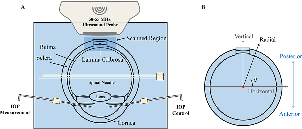
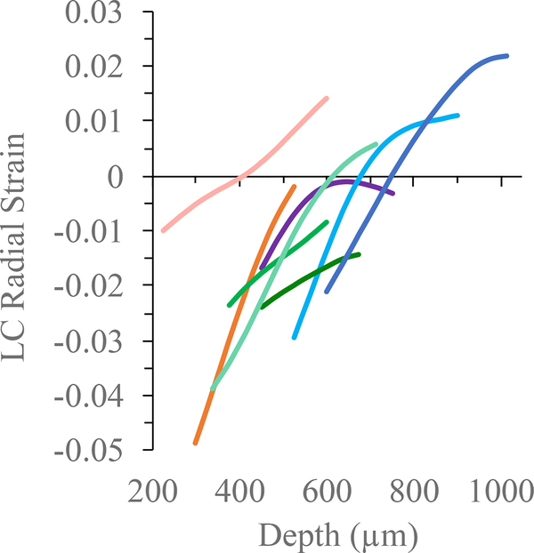
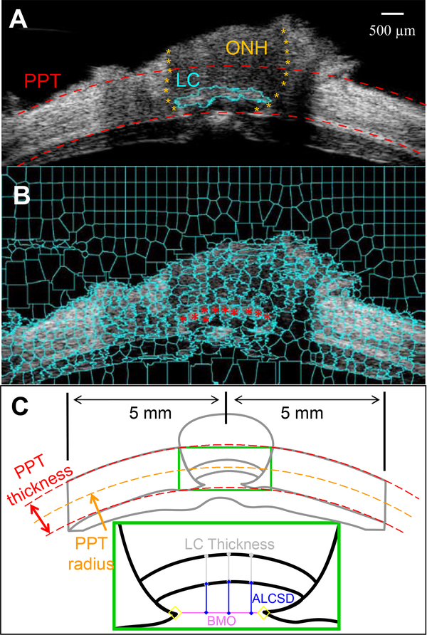
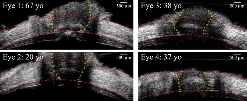
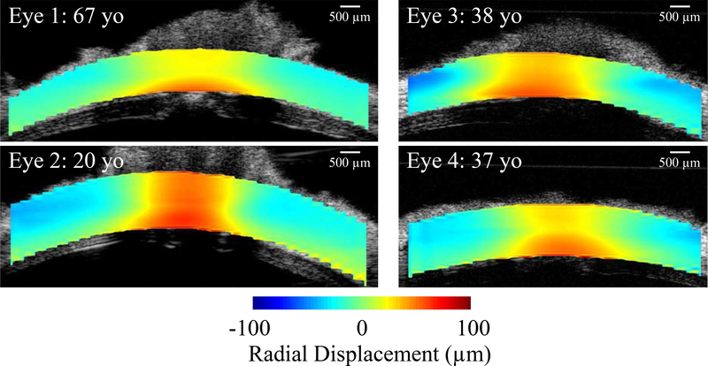
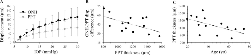
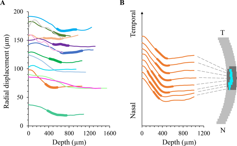
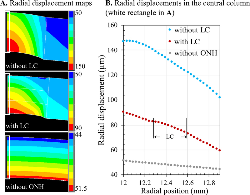
Mechanical insult induced by intraocular pressure (IOP) is likely a driving force in the disease process of glaucoma. This study aimed to evaluate regional displacements in human optic nerve head (ONH) and peripapillary tissue (PPT) in response to acute IOP elevations, and their correlations with morphological characteristics of the posterior eye. Cross-sectional (2D) images of the ONH and PPT in 14 globes of 14 human donors were acquired with high-frequency ultrasound during whole globe inflation from 5 to 30 mm Hg. High-frequency ultrasound has a spatial resolution of tens of micrometers and is capable of imaging through the ONH and PPT thickness. Tissue displacements were calculated using a correlation-based speckle tracking algorithm for a dense matrix of kernels covering the 2D imaging plane. The ONH was manually segmented in the ultrasound B-mode images acquired at 5 mmHg based on echogenicity. The lamina cribrosa (LC) boundaries were visible in eight of the fourteen eyes and the LC region was segmented using a semi-automated superpixel-based method. The ONH had larger radial displacement than the PPT in all tested eyes and the difference increased with increasing IOP. A significant negative correlation was found between ONH-PPT displacement difference and PPT thickness (p < 0.05), while no significant correlations were found between ONH-PPT displacement difference and other morphological parameters including PPT radius of curvature, scleral canal size, LC thickness and anterior LC surface depth. Within the ONH, the radial displacement decreased in the region anterior to and across LC but not in the region posterior to LC. Finite element models using simplified geometry and material properties confirmed the role of LC in reducing the overall ONH radial displacements, but did not predict the displacement gradient change observed experimentally. These results suggested that a thinner PPT may be associated with a larger relative posterior motion of the ONH with respect to the surrounding PPT and the LC may play a major role in preventing excessive posterior displacement of ONH during acute IOP elevations.
眼压(IOP)引起的机械损伤很可能是青光眼疾病进程的驱动力。本研究旨在评估人视神经乳头(ONH)和视盘周围组织(PPT)对急性IOP 升高的区域位移,并评估它们与后眼部形态特征的相关性。在整个眼球从 5 至 30mmHg 充气过程中,使用高频超声获取了 14 个人类供体 14 个眼球的 ONH 和 PPT 的横截面(2D)图像。高频超声的空间分辨率为数十微米,能够穿过 ONH 和 PPT 厚度进行成像。使用基于相关的斑点跟踪算法,对覆盖 2D 成像平面的密集核矩阵进行计算,得出组织位移。在基于回声强度获取的 5mmHg 下的超声 B 模式图像中手动分割 ONH。在 14 只眼睛中,有 8 只眼睛可以看到筛板(LC)边界,并使用半自动基于超像素的方法对 LC 区域进行分割。在所有测试眼,ONH 的径向位移均大于 PPT,且随着 IOP 的增加而增加。ONH-PPT 位移差与 PPT 厚度之间存在显著的负相关(p<0.05),而 ONH-PPT 位移差与其他形态参数(包括 PPT 曲率半径、巩膜管大小、LC 厚度和前 LC 表面深度)之间没有显著相关性。在 ONH 内,LC 前和 LC 处的径向位移减小,但 LC 后区域的位移没有减小。使用简化几何形状和材料特性的有限元模型证实了 LC 在降低整体 ONH 径向位移中的作用,但没有预测到实验中观察到的位移梯度变化。这些结果表明,较薄的 PPT 可能与 ONH 相对于周围 PPT 的较大相对后向运动有关,LC 可能在急性 IOP 升高期间防止 ONH 的过度后向位移方面发挥主要作用。