• 文献检索
  • 文档翻译
  • 深度研究
  • 学术资讯
  • Suppr Zotero 插件Zotero 插件
  • 邀请有礼
  • 套餐&价格
  • 历史记录
应用&插件
Suppr Zotero 插件Zotero 插件浏览器插件Mac 客户端Windows 客户端微信小程序
定价
高级版会员购买积分包购买API积分包
服务
文献检索文档翻译深度研究API 文档MCP 服务
关于我们
关于 Suppr公司介绍联系我们用户协议隐私条款
关注我们

Suppr 超能文献

核心技术专利:CN118964589B侵权必究
粤ICP备2023148730 号-1Suppr @ 2026

文献检索

告别复杂PubMed语法,用中文像聊天一样搜索,搜遍4000万医学文献。AI智能推荐,让科研检索更轻松。

立即免费搜索

文件翻译

保留排版,准确专业,支持PDF/Word/PPT等文件格式,支持 12+语言互译。

免费翻译文档

深度研究

AI帮你快速写综述,25分钟生成高质量综述,智能提取关键信息,辅助科研写作。

立即免费体验

一种高精度、高效率的数字体相关方法,用于从光学相干断层扫描中描述活体视神经头生物力学特性。

A high-accuracy and high-efficiency digital volume correlation method to characterize in-vivo optic nerve head biomechanics from optical coherence tomography.

机构信息

Department of Ophthalmology, University of Pittsburgh, Pittsburgh, PA, United States.

Department of Preventive and Restorative Dental Sciences, UCSF, San Francisco, CA, United States.

出版信息

Acta Biomater. 2022 Apr 15;143:72-86. doi: 10.1016/j.actbio.2022.02.021. Epub 2022 Feb 20.

DOI:10.1016/j.actbio.2022.02.021
PMID:35196556
原文链接:https://pmc.ncbi.nlm.nih.gov/articles/PMC9035111/
Abstract

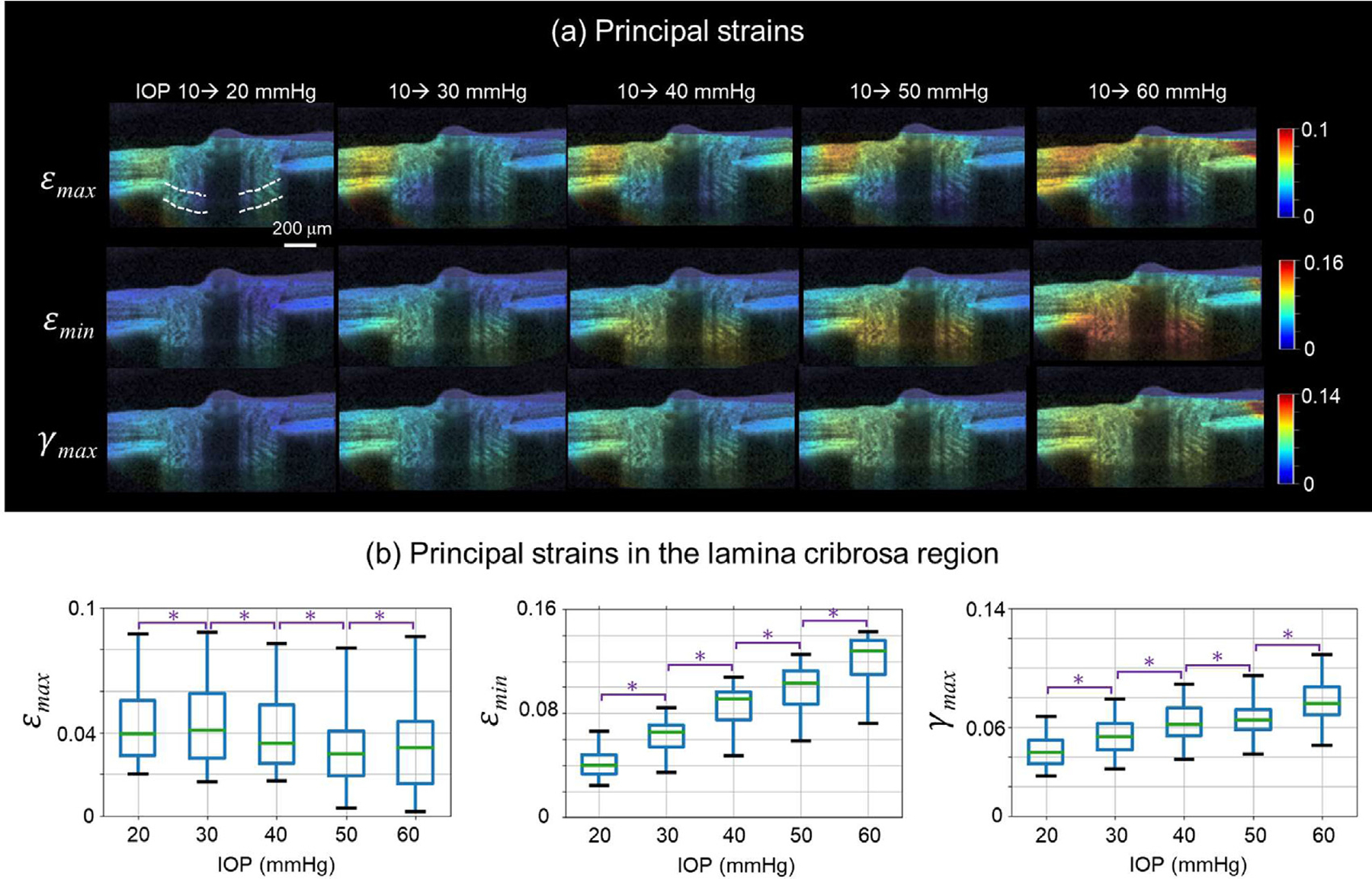
In-vivo optic nerve head (ONH) biomechanics characterization is emerging as a promising way to study eye physiology and pathology. We propose a high-accuracy and high-efficiency digital volume correlation (DVC) method to characterize the in-vivo ONH deformation from optical coherence tomography (OCT) volumes. Using a combination of synthetic tests and analysis of OCTs from monkey ONHs subjected to acutely elevated intraocular pressure, we demonstrate that our proposed methodology overcame several challenges for conventional DVC methods: First, a pre-registration technique was used to remove large ONH rigid body motion in OCT volumes which could lead to analysis failure; second, a modified 3D inverse-compositional Gaussian Newton method was used to ensure sub-voxel accuracy of displacement calculations despite high noise and low image contrast of some OCT volumes; third, a tricubic B-spline interpolation method was applied to improve computational efficiency; fourth, a confidence parameter was introduced to guide the searching path in the displacement calculation; fifth, a confidence-weighted strain calculation method was applied to further improve the accuracy. The proposed DVC method had displacement errors smaller than 0.037 and 0.028 voxels with Gaussian and speckle noises, respectively. The strain errors in the three directions were less than 0.0045 and 0.0018 with Gaussian and speckle noises, respectively. Compared with the conventional DVC method, the proposed method reduced the errors of displacement and strain calculations by up to 70% under large body motions, with 75% lower computation time, while saving about 30% memory. Our study demonstrates the potential of the proposed technique to investigate ONH biomechanics. STATEMENT OF SIGNIFICANCE: The biomechanics of the optic nerve head (ONH) in the posterior pole of the globe play a central role in eye physiology and pathology. The application of digital volume correlation (DVC) to the analysis of optical coherence tomography (OCT) images of the ONH has emerged as a promising way to quantify ONH biomechanics. Conventional DVC methods, however, face several important challenges when analyzing OCT images of the ONH. We introduce a high-accuracy and high-efficiency DVC method to characterize in vivo ONH deformations from OCT volumes. We demonstrate the new method using synthetic tests and actual OCT data from monkey ONHs. The new method also has the potential to be used to study other tissues, as OCT applications continue to expand.

摘要

在体视神经头(ONH)生物力学特征分析作为研究眼睛生理和病理的一种有前途的方法正在出现。我们提出了一种高精度、高效率的数字体相关(DVC)方法,用于从光学相干断层扫描(OCT)体积中描述在体 ONH 变形。通过对受急性眼内压升高影响的猴 ONH 的 OCT 进行综合测试和分析,我们证明了我们提出的方法克服了传统 DVC 方法的几个挑战:首先,使用预配准技术去除 OCT 体积中大的 ONH 刚体运动,这可能导致分析失败;其次,使用改进的三维逆复合高斯牛顿法确保了位移计算的亚像素精度,尽管一些 OCT 体积的噪声和图像对比度低;第三,应用三次 B 样条插值方法提高计算效率;第四,引入置信参数来指导位移计算中的搜索路径;第五,应用置信加权应变计算方法进一步提高精度。所提出的 DVC 方法在具有高斯和散斑噪声的情况下,位移误差分别小于 0.037 和 0.028 体素。三个方向的应变误差分别小于 0.0045 和 0.0018,具有高斯和散斑噪声。与传统的 DVC 方法相比,在大刚体运动下,该方法将位移和应变计算的误差减少了多达 70%,计算时间降低了 75%,同时节省了约 30%的内存。我们的研究表明,该技术具有研究 ONH 生物力学的潜力。

意义声明

眼球后极的视神经头(ONH)的生物力学在眼睛的生理和病理中起着核心作用。数字体相关(DVC)在分析 ONH 的光学相干断层扫描(OCT)图像中的应用已经成为定量 ONH 生物力学的一种有前途的方法。然而,当分析 ONH 的 OCT 图像时,传统的 DVC 方法面临着几个重要的挑战。我们提出了一种高精度、高效率的 DVC 方法,用于从 OCT 体积中描述在体 ONH 变形。我们使用合成测试和来自猴 ONH 的实际 OCT 数据来证明新方法。该新方法也有可能用于研究其他组织,因为 OCT 应用继续扩展。

https://cdn.ncbi.nlm.nih.gov/pmc/blobs/bd4a/9035111/d19cdff5be1c/nihms-1782538-f0012.jpg
https://cdn.ncbi.nlm.nih.gov/pmc/blobs/bd4a/9035111/7a9cde695f88/nihms-1782538-f0001.jpg
https://cdn.ncbi.nlm.nih.gov/pmc/blobs/bd4a/9035111/edefb949017a/nihms-1782538-f0002.jpg
https://cdn.ncbi.nlm.nih.gov/pmc/blobs/bd4a/9035111/3d7c9db5c308/nihms-1782538-f0003.jpg
https://cdn.ncbi.nlm.nih.gov/pmc/blobs/bd4a/9035111/ca97c8385093/nihms-1782538-f0004.jpg
https://cdn.ncbi.nlm.nih.gov/pmc/blobs/bd4a/9035111/d18d2dd677d1/nihms-1782538-f0005.jpg
https://cdn.ncbi.nlm.nih.gov/pmc/blobs/bd4a/9035111/4a24c0a5405a/nihms-1782538-f0006.jpg
https://cdn.ncbi.nlm.nih.gov/pmc/blobs/bd4a/9035111/ca26a481e817/nihms-1782538-f0007.jpg
https://cdn.ncbi.nlm.nih.gov/pmc/blobs/bd4a/9035111/14609b403f33/nihms-1782538-f0008.jpg
https://cdn.ncbi.nlm.nih.gov/pmc/blobs/bd4a/9035111/f90917c83ffb/nihms-1782538-f0009.jpg
https://cdn.ncbi.nlm.nih.gov/pmc/blobs/bd4a/9035111/155687f1101f/nihms-1782538-f0010.jpg
https://cdn.ncbi.nlm.nih.gov/pmc/blobs/bd4a/9035111/fe0c35a353fd/nihms-1782538-f0011.jpg
https://cdn.ncbi.nlm.nih.gov/pmc/blobs/bd4a/9035111/d19cdff5be1c/nihms-1782538-f0012.jpg

相似文献

1
A high-accuracy and high-efficiency digital volume correlation method to characterize in-vivo optic nerve head biomechanics from optical coherence tomography.一种高精度、高效率的数字体相关方法,用于从光学相干断层扫描中描述活体视神经头生物力学特性。
Acta Biomater. 2022 Apr 15;143:72-86. doi: 10.1016/j.actbio.2022.02.021. Epub 2022 Feb 20.
2
Verification of a virtual fields method to extract the mechanical properties of human optic nerve head tissues in vivo.一种用于在体内提取人视神经乳头组织力学特性的虚拟场方法的验证。
Biomech Model Mechanobiol. 2017 Jun;16(3):871-887. doi: 10.1007/s10237-016-0858-2. Epub 2016 Dec 1.
3
In vivo optic nerve head biomechanics: performance testing of a three-dimensional tracking algorithm.活体视神经头生物力学:三维跟踪算法的性能测试。
J R Soc Interface. 2013 Jul 24;10(87):20130459. doi: 10.1098/rsif.2013.0459. Print 2013 Oct 6.
4
In Vivo 3-Dimensional Strain Mapping of the Optic Nerve Head Following Intraocular Pressure Lowering by Trabeculectomy.小梁切除术后眼压降低对眼内压降低对视乳头的三维应变的影响。
Ophthalmology. 2016 Jun;123(6):1190-200. doi: 10.1016/j.ophtha.2016.02.008. Epub 2016 Mar 16.
5
In vivo characterization of the deformation of the human optic nerve head using optical coherence tomography and digital volume correlation.应用光相干断层扫描和数字体相关技术对人视神经乳头变形的体内特征进行研究。
Acta Biomater. 2019 Sep 15;96:385-399. doi: 10.1016/j.actbio.2019.06.050. Epub 2019 Jul 3.
6
AI-based clinical assessment of optic nerve head robustness superseding biomechanical testing.基于人工智能的视神经头稳健性临床评估取代生物力学测试。
Br J Ophthalmol. 2024 Jan 29;108(2):223-231. doi: 10.1136/bjo-2022-322374.
7
A method to estimate biomechanics and mechanical properties of optic nerve head tissues from parameters measurable using optical coherence tomography.一种从使用光学相干断层扫描可测量的参数来估计视神经乳头组织生物力学和力学特性的方法。
IEEE Trans Med Imaging. 2014 Jun;33(6):1381-9. doi: 10.1109/TMI.2014.2312133. Epub 2014 Mar 14.
8
Three-dimensional surface presentation of optic nerve head from SPECTRALIS OCT images: observing glaucoma patients.基于SPECTRALIS OCT图像的视神经乳头三维表面呈现:青光眼患者观察
Int Ophthalmol. 2019 Sep;39(9):1939-1947. doi: 10.1007/s10792-018-1023-y. Epub 2018 Sep 24.
9
Computational study on the effects of central retinal blood vessels with asymmetric geometries on optic nerve head biomechanics.基于中央视网膜血管非对称结构对视神经头生物力学影响的计算研究。
Med Eng Phys. 2024 Jan;123:104086. doi: 10.1016/j.medengphy.2023.104086. Epub 2023 Dec 12.
10
High-accuracy optical coherence elastography digital volume correlation methods to measure depth regions with low correlation.高精度光学相干弹性成像数字体相关方法测量低相关深度区域。
J Biophotonics. 2024 Jan;17(1):e202300094. doi: 10.1002/jbio.202300094. Epub 2023 Oct 9.

引用本文的文献

1
Research progress of in vivo measurement methods of myopia sclera biomechanics.近视巩膜生物力学体内测量方法的研究进展
Adv Ophthalmol Pract Res. 2025 Jun 14;5(3):205-211. doi: 10.1016/j.aopr.2025.04.006. eCollection 2025 Aug-Sep.
2
Impact of Elevated Intraocular Pressure on Lamina Cribrosa Oxygenation: A Combined Experimental-Computational Study on Monkeys.眼压升高对筛板氧合的影响:一项对猴子的实验与计算相结合的研究
Ophthalmol Sci. 2025 Jan 31;5(3):100725. doi: 10.1016/j.xops.2025.100725. eCollection 2025 May-Jun.
3
Biomechanical Strain Responses in the Sclera, Choroid, and Retina in Glaucoma Patients After Intraocular Pressure Lowering.

本文引用的文献

1
Histologic validation of optical coherence tomography-based three-dimensional morphometric measurements of the human optic nerve head: Methodology and preliminary results.基于光学相干断层扫描的人视神经头三维形态测量的组织学验证:方法学和初步结果。
Exp Eye Res. 2021 Apr;205:108475. doi: 10.1016/j.exer.2021.108475. Epub 2021 Jan 28.
2
Biomechanics of the optic nerve head and peripapillary sclera in a mouse model of glaucoma.青光眼小鼠模型中视神经乳头和视乳头周围巩膜的生物力学
J R Soc Interface. 2020 Dec;17(173):20200708. doi: 10.1098/rsif.2020.0708. Epub 2020 Dec 16.
3
IOP-induced regional displacements in the optic nerve head and correlation with peripapillary sclera thickness.
青光眼患者眼压降低后巩膜、脉络膜和视网膜的生物力学应变反应
J Biomech Eng. 2025 May 1;147(5). doi: 10.1115/1.4068155.
4
Proposing a Methodology for Axon-Centric Analysis of IOP-Induced Mechanical Insult.提出一种针对眼压诱导的机械损伤的轴突中心分析方法。
Invest Ophthalmol Vis Sci. 2024 Nov 4;65(13):1. doi: 10.1167/iovs.65.13.1.
5
Impact of elevated IOP on lamina cribrosa oxygenation; A combined experimental-computational study on monkeys.眼压升高对筛板氧合的影响;一项针对猴子的实验与计算相结合的研究
bioRxiv. 2024 Sep 10:2024.09.05.609208. doi: 10.1101/2024.09.05.609208.
6
The Robust Lamina Cribrosa Vasculature: Perfusion and Oxygenation Under Elevated Intraocular Pressure.稳健的神经节细胞层血管:高眼压下的灌注和氧合。
Invest Ophthalmol Vis Sci. 2024 May 1;65(5):1. doi: 10.1167/iovs.65.5.1.
7
Denoising OCT videos based on temporal redundancy.基于时间冗余的 OCT 视频去噪。
Sci Rep. 2024 Mar 19;14(1):6605. doi: 10.1038/s41598-024-56935-0.
8
Reverse translation of artificial intelligence in glaucoma: Connecting basic science with clinical applications.青光眼人工智能的反向翻译:连接基础科学与临床应用。
Front Ophthalmol (Lausanne). 2023;2. doi: 10.3389/fopht.2022.1057896. Epub 2023 Jan 4.
9
Optical Coherence Tomography Angiography Demonstrates Strain and Volume Effects on Optic Disk and Peripapillary Vasculature Caused by Horizontal Duction.光学相干断层扫描血管造影显示水平直肌牵拉对视盘和视盘周围血管的应变和容积效应。
Curr Eye Res. 2023 May;48(5):518-527. doi: 10.1080/02713683.2023.2172185. Epub 2023 Feb 27.
10
Comparing Acute IOP-Induced Lamina Cribrosa Deformations Premortem and Postmortem.比较生前和死后急性眼压诱导的筛板变形。
Transl Vis Sci Technol. 2022 Dec 1;11(12):1. doi: 10.1167/tvst.11.12.1.
IOP 诱导的视神经头区域位移与视盘周围巩膜厚度的相关性。
Exp Eye Res. 2020 Nov;200:108202. doi: 10.1016/j.exer.2020.108202. Epub 2020 Aug 27.
4
Lamina Cribrosa Moves Anteriorly After Trabeculectomy in Myopic Eyes.近视眼行小梁切除术后,筛板向前移动。
Invest Ophthalmol Vis Sci. 2020 Jun 3;61(6):36. doi: 10.1167/iovs.61.6.36.
5
The inflation response of the human lamina cribrosa and sclera: Analysis of deformation and interaction.人颅筛骨和巩膜的膨胀反应:变形与相互作用分析。
Acta Biomater. 2020 Apr 1;106:225-241. doi: 10.1016/j.actbio.2020.01.049. Epub 2020 Feb 8.
6
Characterization of chemoelastic effects in arteries using digital volume correlation and optical coherence tomography.采用数字体积相关技术和光学相干断层扫描技术对动脉的化学弹性效应进行表征。
Acta Biomater. 2020 Jan 15;102:127-137. doi: 10.1016/j.actbio.2019.11.049. Epub 2019 Nov 30.
7
Relationship of Corneal Hysteresis and Anterior Lamina Cribrosa Displacement in Glaucoma.青光眼患者角膜滞后与前房梁板位移的关系。
Am J Ophthalmol. 2020 Apr;212:134-143. doi: 10.1016/j.ajo.2019.11.017. Epub 2019 Nov 23.
8
Racial Differences in the Association of Anterior Lamina Cribrosa Surface Depth and Glaucoma Severity in the African Descent and Glaucoma Evaluation Study (ADAGES).非裔美国人与青光眼评估研究(ADAGES)中前筛板表面深度与青光眼严重程度的关联存在种族差异。
Invest Ophthalmol Vis Sci. 2019 Oct 1;60(13):4496-4502. doi: 10.1167/iovs.19-26645.
9
In vivo characterization of the deformation of the human optic nerve head using optical coherence tomography and digital volume correlation.应用光相干断层扫描和数字体相关技术对人视神经乳头变形的体内特征进行研究。
Acta Biomater. 2019 Sep 15;96:385-399. doi: 10.1016/j.actbio.2019.06.050. Epub 2019 Jul 3.
10
Synchrotron tomography of intervertebral disc deformation quantified by digital volume correlation reveals microstructural influence on strain patterns.基于数字体图像相关技术的椎间盘变形同步辐射断层成像定量分析显示了微观结构对变形模式的影响。
Acta Biomater. 2019 Jul 1;92:290-304. doi: 10.1016/j.actbio.2019.05.021. Epub 2019 May 11.