Sabbaghi Hamideh, Ahmadieh Hamid, Jalili Jalil, Behnaz Nazanin, Fakhri Maryam, Suri Fatemeh, Kheiri Bahareh, Rajabpour Mojtaba, Entezari Morteza, Daftarian Narsis
Ophthalmic Epidemiology Research Center, Research Institute for Ophthalmology and Vision Science, Shahid Beheshti University of Medical Sciences, Tehran, Iran.
Department of Optometry, School of Rehabilitation, Shahid Beheshti University of Medical Sciences, Tehran, Iran.
J Ophthalmic Vis Res. 2020 Aug 6;15(3):351-361. doi: 10.18502/jovr.v15i3.7454. eCollection 2020 Jul-Sep.
To compare the choroidal thickness among eyes with retinitis pigmentosa (RP), Stargardt disease, Usher syndrome, cone-rod dystrophy, and healthy eyes of sex- and age-matched individuals.
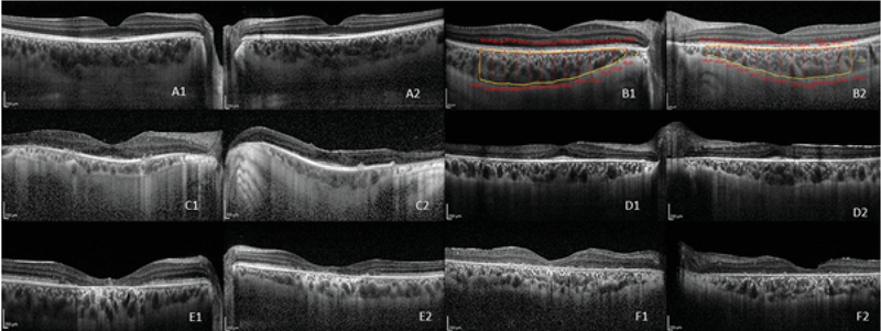
In this comparative study, 503 eyes with RP ( = 264), cone-rod dystrophy ( = 109), Stargardt disease ( = 76), and Usher syndrome ( = 54) were included. To validate the data, 109 healthy eyes of 56 sex- and age-matched individuals were studied as controls. Choroidal imaging was performed using enhanced depth imaging-optical coherence tomography. Choroidal thickness was measured manually using MATLAB software at 13 points in nasal and temporal directions from the foveal center with the interval of 500 µm and the choroidal area encompassing the measured points was calculated automatically.
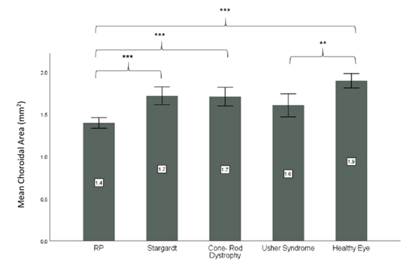
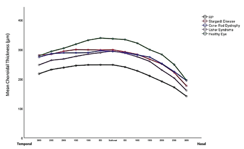
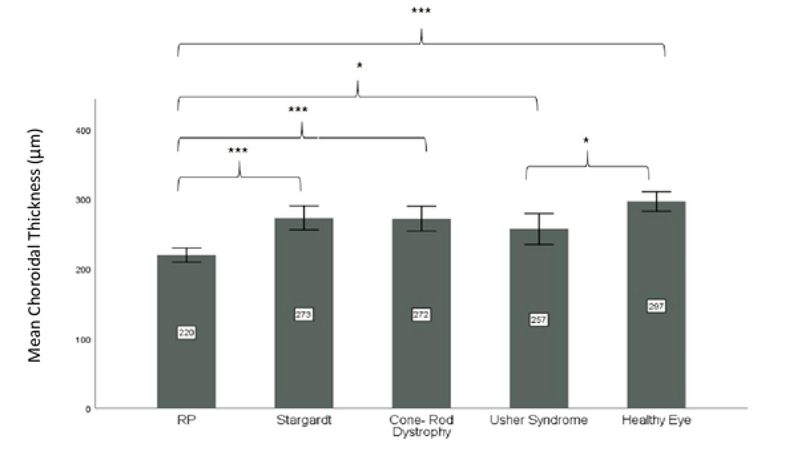
The mean age was 36.33 13.07 years (range, 5 to 72 years). The mean choroidal thickness at 13 points of the control eyes was statistically significantly higher than that in eyes with RP ( 0.001) and Usher syndrome ( 0.05), but not significantly different from that in eyes with Stargardt disease and cone-rod dystrophy. Among different inherited retinal dystrophies (IRDs), the choroidal thickness was the lowest in eyes with RP ( 0.001). Choroidal thickness in the subfoveal area correlated negatively with best-corrected visual acuity ( = 0.264, 0.001) and the duration of ocular symptoms ( = 0.341, 0.001) in all studied IRDs. No significant correlation was observed between the subfoveal choroidal thickness and central macular thickness ( = 0.24, = 0.576).
Choroidal thinning in four different types of IRDs does not follow a similar pattern and depends on the type of IRD and the duration of ocular symptoms. A larger cohort is required to verify these findings.
比较视网膜色素变性(RP)、斯塔加特病、Usher综合征、锥杆营养不良患者的眼睛与性别和年龄匹配的健康人眼睛的脉络膜厚度。
在这项比较研究中,纳入了503只患有RP(n = 264)、锥杆营养不良(n = 109)、斯塔加特病(n = 76)和Usher综合征(n = 54)的眼睛。为了验证数据,对56名性别和年龄匹配个体的109只健康眼睛进行了研究作为对照。使用增强深度成像光学相干断层扫描进行脉络膜成像。使用MATLAB软件在距黄斑中心鼻侧和颞侧方向的13个点手动测量脉络膜厚度,间隔为500 µm,并自动计算包含测量点的脉络膜面积。
平均年龄为36.33±13.07岁(范围为5至72岁)。对照眼13个点的平均脉络膜厚度在统计学上显著高于RP患者的眼睛(P < 0.001)和Usher综合征患者的眼睛(P < 0.05),但与斯塔加特病和锥杆营养不良患者的眼睛无显著差异。在不同的遗传性视网膜营养不良(IRD)中,RP患者眼睛的脉络膜厚度最低(P < 0.001)。在所有研究的IRD中,黄斑下区域的脉络膜厚度与最佳矫正视力呈负相关(r = -0.264,P < 0.001)和眼部症状持续时间呈负相关(r = -0.341,P < 0.001)。未观察到黄斑下脉络膜厚度与中心黄斑厚度之间存在显著相关性(r = 0.24,P = 0.576)。
四种不同类型IRD中的脉络膜变薄模式不同,取决于IRD的类型和眼部症状的持续时间。需要更大的队列来验证这些发现。