Nuclear Medicine and PET, Department of Surgical Sciences, Uppsala University, SE-751 85 Uppsala, Sweden; Medical Physics, Uppsala University Hospital, SE-751 85 Uppsala, Sweden.
Radiation Physics, Skåne University Hospital, SE-221 85 Lund, Sweden.
Neuroimage Clin. 2020;28:102386. doi: 10.1016/j.nicl.2020.102386. Epub 2020 Aug 19.
To assess how some of the new developments in brain positron emission tomography (PET) image reconstruction affect quantitative measures and software-aided assessment of pathology in patients with neurodegenerative diseases.
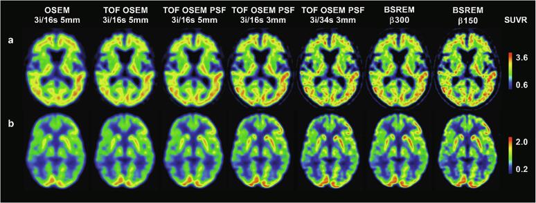
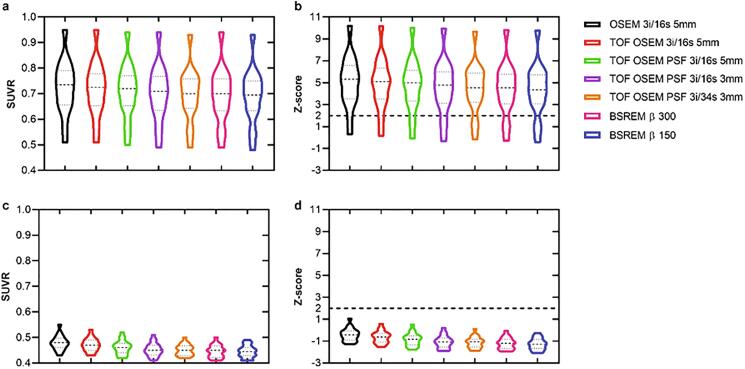
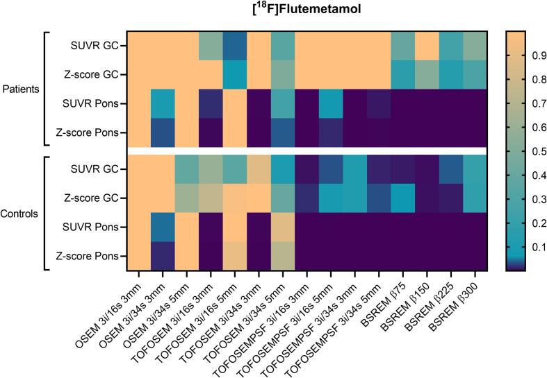
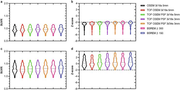
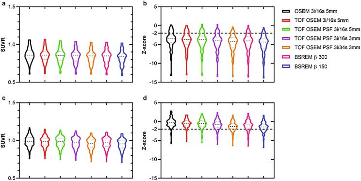
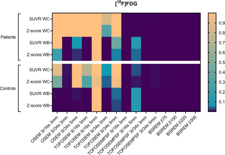
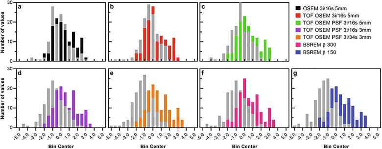
PET data were grouped into four cohorts: prodromal Alzheimer's disease patients and controls receiving [F]flutemetamol, and neurodegenerative disease patients and controls receiving [F]FDG PET scans. Reconstructed images were obtained by ordered-subsets expectation maximization (OSEM; 3 iterations (i), 16/34 subsets (s), 3/5-mm filter, ±time-of-flight (TOF), ±point-spread function (PSF)) and block-sequential regularized expectation maximization (BSREM; TOF, PSF, β-value 75-300). Standardized uptake value ratios (SUVR) and z-scores were calculated (CortexID Suite, GE Healthcare) using cerebellar gray matter, pons, whole cerebellum and whole brain as reference regions.
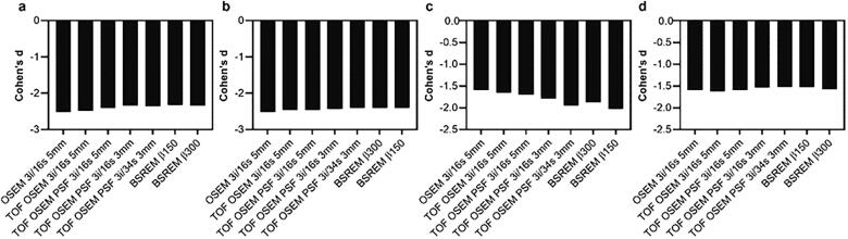
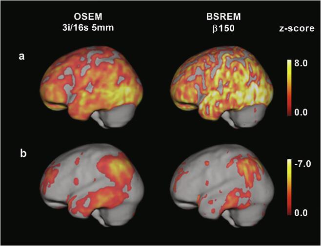
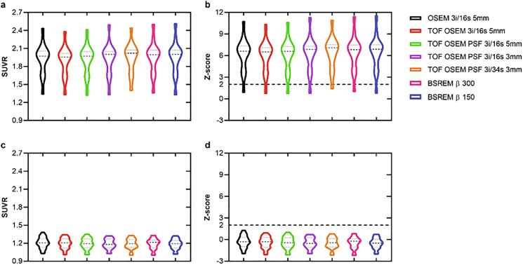
In controls, comparable results to the normal database were obtained with OSEM 3i/16 s 5-mm reconstruction. TOF, PSF and BSREM either increased or decreased the relative uptake difference to the normal subjects' database within the software, depending on the tracer and chosen reference area, i.e. resulting in increased absolute z-scores. Normalizing to pons and whole brain for [F]flutemetamol and [F]FDG, respectively, increased absolute differences between reconstructions methods compared to normalizing to cerebellar gray matter and whole cerebellum when applying TOF, PSF and BSREM.
Software-aided assessment of patient pathologies should be used with caution when employing other image reconstruction methods than those used for acquisition of the normal database.
评估脑正电子发射断层扫描(PET)图像重建的一些新进展如何影响神经退行性疾病患者的定量测量和软件辅助病理评估。
将 PET 数据分为四组:前驱性阿尔茨海默病患者和接受 [F]flutemetamol 的对照组,以及接受 [F]FDG PET 扫描的神经退行性疾病患者和对照组。通过有序子集期望最大化(OSEM;3 次迭代(i)、16/34 子集(s)、3/5mm 滤波器、±时间-of-flight(TOF)、±点扩散函数(PSF))和块顺序正则化期望最大化(BSREM;TOF、PSF、β 值 75-300)获得重建图像。使用小脑灰质、脑桥、全小脑和全脑作为参考区域,使用 CortexID 套件(通用电气医疗)计算标准化摄取值比(SUVr)和 z 分数。
在对照组中,OSEM 3i/16s 5mm 重建获得与正常数据库相当的结果。TOF、PSF 和 BSREM 要么增加,要么减少了与软件中正常受试者数据库的相对摄取差异,这取决于示踪剂和所选的参考区域,即导致绝对 z 分数增加。分别为 [F]flutemetamol 和 [F]FDG 归一化为脑桥和全脑,与将 TOF、PSF 和 BSREM 应用于小脑灰质和全小脑相比,重建方法之间的绝对差异增加。
在使用比正常数据库采集方法不同的图像重建方法时,应谨慎使用软件辅助评估患者的病理。