He Ming-Qian, Wang Jing-Ya, Wang Yue, Sui Jing, Zhang Meng, Ding Xi, Zhao Yang, Chen Zi-Yi, Ren Xiao-Xiao, Shi Bing-Yin
Department of Endocrinology, The First Affiliated Hospital of Xi'an JiaoTong University, Xi'an, Shaanxi 710061, China.
Department of Endocrinology and International Medical Center, The First Affiliated Hospital of Xi'an JiaoTong University, Xi'an, Shaanxi 710061, China.
Chronic Dis Transl Med. 2020 Jul 18;6(3):198-207. doi: 10.1016/j.cdtm.2020.06.003. eCollection 2020 Sep.
To date, there is only scare evidence characterizing the temporal features and progression of metabolic dysfunction in high-fat diet (HFD)-fed obese mice. Hence, its specific pathogenesis remains unclear.
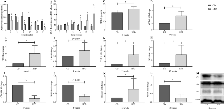
Sixty 6-week-old male C57BL/6J mice were randomly divided into HFD and control diet (CD) groups and sacrificed at 1, 5, 9, 13, 17, and 21 weeks, respectively. At weekly intervals, intraperitoneal glucose tolerance testing (IPGTT) and intraperitoneal insulin tolerance testing (IPITT) were performed in both groups. A detailed time course in HFD-fed mice was investigated by evaluating the initiation of glucose homeostasis impairment, dyslipidemia, systemic insulin sensitivity, monocyte chemoattractant protein-1 (MCP-1) levels, epididymal white adipose tissue (eWAT) expansion, macrophage content changes, pro-inflammatory (M1)/anti-inflammatory (M2) macrophage imbalance, lipid accumulation in the liver, and β-cell morphometry in the pancreas.
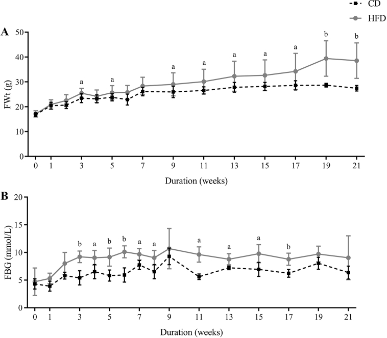
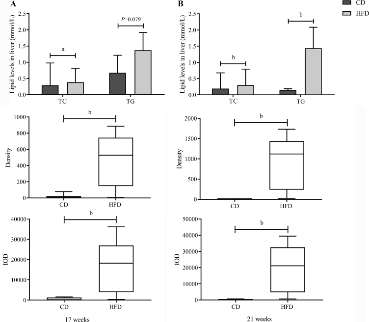
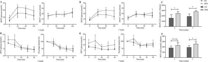
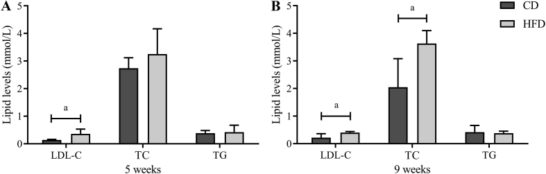
In the HFD group, progressive weight gain and impairments in glucose metabolism (elevated fasting blood glucose and area under the curve (AUC) of IPGTT) were observed from the 3rd week, and a significantly elevated AUC of IPITT was first detected after week 7 of HFD feeding. As for dyslipidemia, after 9 weeks of feeding, the low-density lipoprotein cholesterol level and total cholesterol level in HFD group were significantly higher than those in the CD group (all < 0.05), whereas no significant differences were shown in triglyceride level. Adipocyte size increased significantly in the HFD group in the 1st week, a phenotypic switch in eWAT from anti-inflammatory (M2) to pro-inflammatory (M1) macrophages was observed in the 5th week, and the metabolic inflammation was distinct in eWAT in the 9th week. Additionally, liver steatosis was considerably obvious at the 17th week and pancreatic β-cell morphometry did not change during 21 weeks of HFD feeding.
The eWAT expansion was detected early in HFD-induced obese mice, which occurred prior to obvious insulin resistance.
迄今为止,仅有少量证据描述高脂饮食(HFD)喂养的肥胖小鼠代谢功能障碍的时间特征和进展情况。因此,其具体发病机制仍不清楚。
将60只6周龄雄性C57BL/6J小鼠随机分为高脂饮食组和对照饮食(CD)组,分别在第1、5、9、13、17和21周处死。两组每周进行一次腹腔葡萄糖耐量试验(IPGTT)和腹腔胰岛素耐量试验(IPITT)。通过评估葡萄糖稳态损害的起始、血脂异常、全身胰岛素敏感性、单核细胞趋化蛋白-1(MCP-1)水平、附睾白色脂肪组织(eWAT)扩张、巨噬细胞含量变化、促炎(M1)/抗炎(M2)巨噬细胞失衡、肝脏脂质蓄积以及胰腺β细胞形态计量学,研究高脂饮食喂养小鼠的详细时间进程。
在高脂饮食组中,从第3周开始观察到体重逐渐增加和葡萄糖代谢受损(空腹血糖升高和IPGTT曲线下面积(AUC)升高),在高脂饮食喂养7周后首次检测到IPITT的AUC显著升高。至于血脂异常,喂养9周后,高脂饮食组的低密度脂蛋白胆固醇水平和总胆固醇水平显著高于对照饮食组(均P<0.05),而甘油三酯水平无显著差异。高脂饮食组在第1周脂肪细胞大小显著增加,在第5周观察到eWAT中巨噬细胞从抗炎(M2)向促炎(M1)的表型转变,在第9周eWAT中的代谢炎症明显。此外,肝脂肪变性在第17周相当明显,在高脂饮食喂养21周期间胰腺β细胞形态计量学未发生变化。
在高脂饮食诱导的肥胖小鼠中,eWAT扩张在早期即可检测到,且发生在明显的胰岛素抵抗之前。