Department of Ophthalmology, Leiden University Medical Center, Leiden, The Netherlands.
University Neurosurgical Centre Holland, Leiden University Medical Centre, Haaglanden Medical Centre and Haga Teaching Hospital, Leiden and The Hague, The Netherlands.
Acta Neurochir (Wien). 2021 Jan;163(1):73-82. doi: 10.1007/s00701-020-04554-9. Epub 2020 Sep 4.
BACKGROUND: Most spheno-orbital meningioma series span multiple decades, and predictors of visual outcomes have not yet been systemically assessed. We describe visual outcomes in a recent cohort and assess predictors of postoperative visual outcomes.
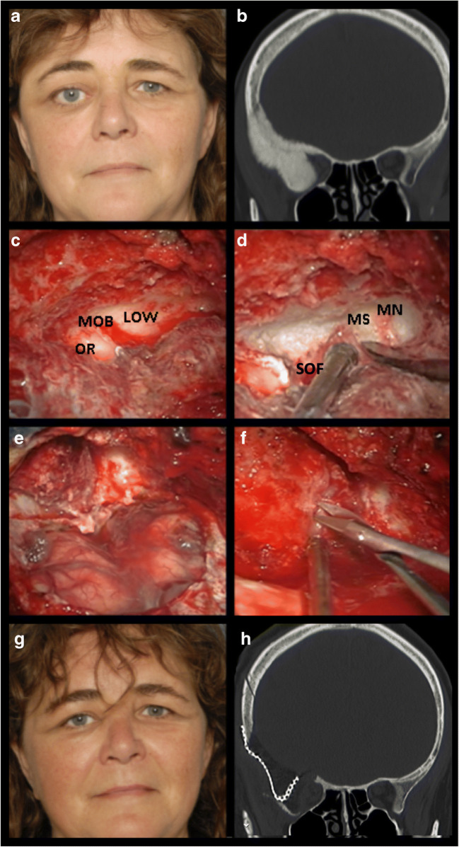
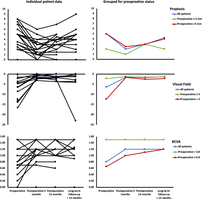
Consecutive case series operated by a team of a neurosurgeon and orbital surgeon between May 2015 and January 2019. Best corrected visual acuity (BCVA), visual fields (static perimetry), and relative proptosis were measured preoperatively and postoperatively at 3/6/12 months after which it was assessed yearly. Predictors were assessed with linear regression analysis.
Nineteen patients (all WHO grade I) were operated by the pterional approach (median follow-up 2.4 years). Preoperative visual acuity deficits (n = 10) normalized in 70% and improved in 10% (median preoperative: 0.8, postoperative: 1.2, p = 0.021). Preoperative visual field deficits (n = 8) normalized in all patients (preoperative: - 6.5 dB, postoperative: - 1.5 dB, p = 0.008). Preoperative proptosis (n = 16) normalized in 44% and improved in 56% (preoperative: 5 mm, postoperative: 2 mm, p < 0.001). BCVA and visual fields remained stable at longer follow-up in 95% of patients, while 21% showed progression of proptosis. Predictors for worse longer-term (> 12 months) BCVA were worse preoperative BCVA (p = 0.002) and diagnosis of multiple meningioma (p = 0.021). Predictors for worse longer-term visual fields were higher diameter of hyperostosis (p = 0.009) and higher Simpson grade (p = 0.032). Predictor for short-term (3 months) proptosis was preoperative proptosis (p = 0.006).
We recommend surgery, even of patients with minimal visual impairment or hyperostosis, as patients who present with deteriorated visual function or extensive hyperostosis are less likely to have postoperative visual outcomes restored to normal.
背景:大多数蝶眶脑膜瘤系列跨越多个十年,视觉结果的预测因素尚未系统评估。我们描述了最近一组患者的视觉结果,并评估了术后视觉结果的预测因素。
连续病例系列,由神经外科医生和眼眶外科医生团队于 2015 年 5 月至 2019 年 1 月进行手术。在术后 3、6、12 个月以及之后每年测量最佳矫正视力(BCVA)、视野(静态视野计)和相对眼球突出度。使用线性回归分析评估预测因素。
19 名(均为 WHO 分级 I)患者通过翼点入路进行手术(中位随访 2.4 年)。术前视力障碍(n=10)中有 70%恢复正常,10%改善(术前中位值:0.8,术后:1.2,p=0.021)。术前视野缺损(n=8)在所有患者中均恢复正常(术前:-6.5dB,术后:-1.5dB,p=0.008)。术前眼球突出(n=16)有 44%恢复正常,56%改善(术前:5mm,术后:2mm,p<0.001)。95%的患者在更长的随访时间内 BCVA 和视野保持稳定,而 21%的患者出现眼球突出进展。术后 12 个月以上(>12 个月)BCVA 较差的预测因素为术前 BCVA 较差(p=0.002)和多发性脑膜瘤诊断(p=0.021)。术后 12 个月以上视野较差的预测因素为骨肥厚直径较大(p=0.009)和 Simpson 分级较高(p=0.032)。短期(3 个月)眼球突出的预测因素是术前眼球突出(p=0.006)。
我们建议手术治疗,即使是对视力障碍或骨肥厚最小的患者,因为出现视觉功能恶化或广泛骨肥厚的患者术后视觉结果恢复正常的可能性较小。