Heidinger Benedikt H, Kifjak Daria, Prayer Florian, Beer Lucian, Milos Ruxandra-Iulia, Röhrich Sebastian, Arndt Hanka, Prosch Helmut
Universitätsklinik für Radiologie und Nuklearmedizin, Medizinische Universität Wien, Währinger Gürtel 18-20, 1090, Wien, Österreich.
Institut für Diagnostische und Interventionelle Radiologie, Kinder- und Neuroradiologie, Universitätsmedizin Rostock, Rostock, Deutschland.
Radiologe. 2020 Oct;60(10):908-915. doi: 10.1007/s00117-020-00749-4.
Since its emergence in late 2019, the disease caused by the novel coronavirus, termed COVID-19, has been declared a pandemic by the World Health Organization. Reference standard for the diagnosis of COVID-19 is a positive reverse transcription polymerase chain reaction (RT-PCR) test. While the RT-PCR shows a high specificity, its sensitivity depends on the duration of symptoms, viral load, quality of the sample, and the assay used.
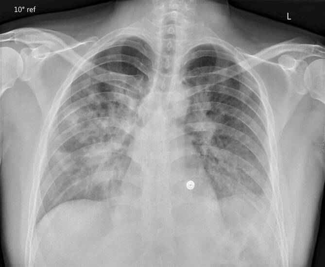
Chest radiography and computed tomography (CT) of the chest are the imaging modalities primarily used for assessment of the lung manifestations, extent, and complications of COVID-19 pneumonia.
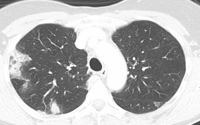
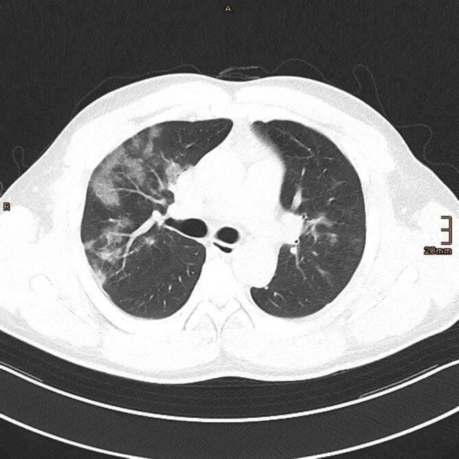
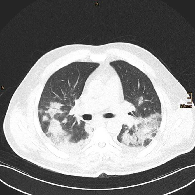
Sensitivity and specificity of chest radiography is low. While sensitivity of CT for detecting COVID-19 pneumonia is high-averaging around 90%-its specificity is low-between 25 and 33%.
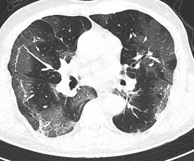
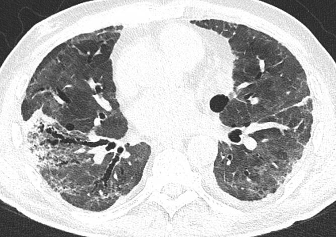
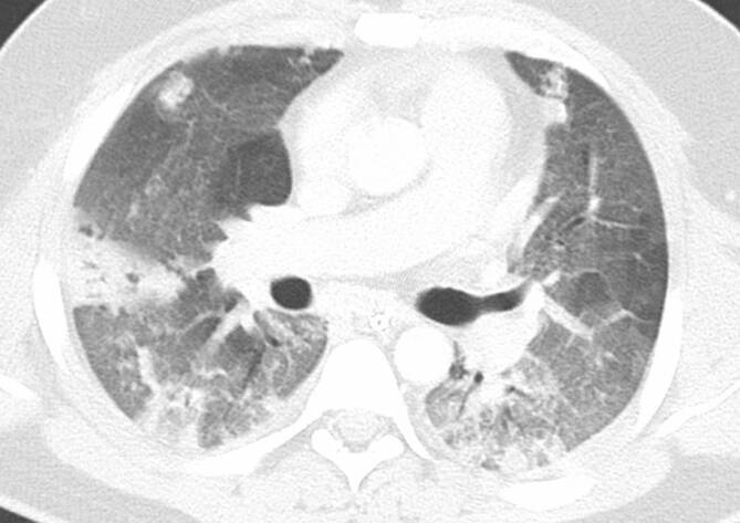
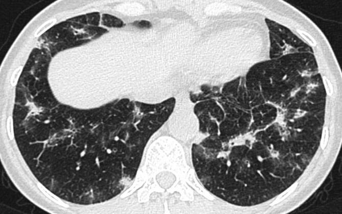
Indications for imaging in patients with suspected or diagnosed COVID-19 infection should be carefully considered to minimize the risk of infection for medical personnel and other patients. Imaging, particularly CT, can assess disease extent, complications, and differential diagnoses. COVID-19 pneumonia typically presents with bilateral, subpleural areas of ground glass opacifications with or without consolidations. During the course of the disease features resembling organizing pneumonia can occur. Follow-up examinations after recovery from COVID-19 pneumonia should focus on fibrotic changes of the lung parenchyma.
自2019年末出现以来,由新型冠状病毒引起的疾病(称为COVID-19)已被世界卫生组织宣布为大流行病。COVID-19诊断的参考标准是逆转录聚合酶链反应(RT-PCR)检测呈阳性。虽然RT-PCR具有高特异性,但其敏感性取决于症状持续时间、病毒载量、样本质量和所使用的检测方法。
胸部X线摄影和胸部计算机断层扫描(CT)是主要用于评估COVID-19肺炎的肺部表现、范围和并发症的成像方式。
胸部X线摄影的敏感性和特异性较低。虽然CT检测COVID-19肺炎的敏感性较高——平均约为90%——但其特异性较低,在25%至33%之间。
对于疑似或确诊COVID-19感染的患者,应仔细考虑成像的指征,以尽量减少医务人员和其他患者的感染风险。成像,尤其是CT,可以评估疾病范围、并发症和鉴别诊断。COVID-19肺炎通常表现为双侧胸膜下磨玻璃样混浊区域,可伴有或不伴有实变。在疾病过程中,可出现类似机化性肺炎的特征。COVID-19肺炎康复后的随访检查应侧重于肺实质的纤维化改变。