Department of Biomedical Engineering, University of Delaware, Newark, Delaware, USA.
Thayer School of Engineering, Dartmouth College, Hanover, New Hampshire, USA.
Hum Brain Mapp. 2020 Dec 15;41(18):5282-5300. doi: 10.1002/hbm.25192. Epub 2020 Sep 15.
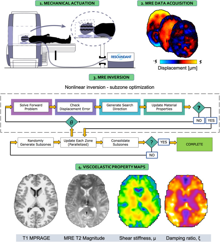
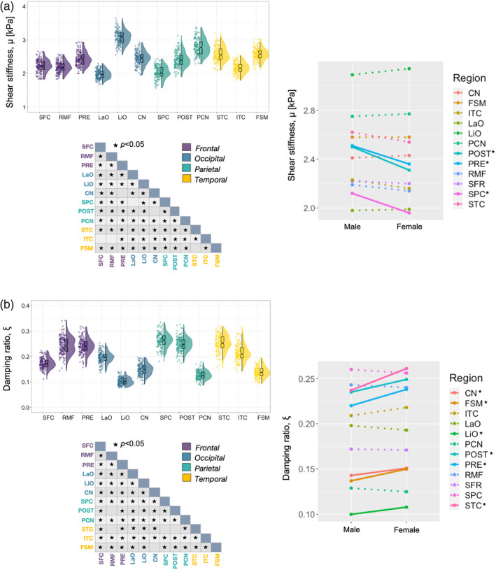
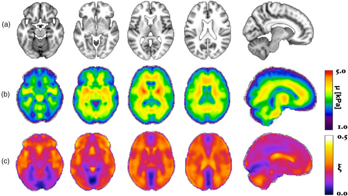
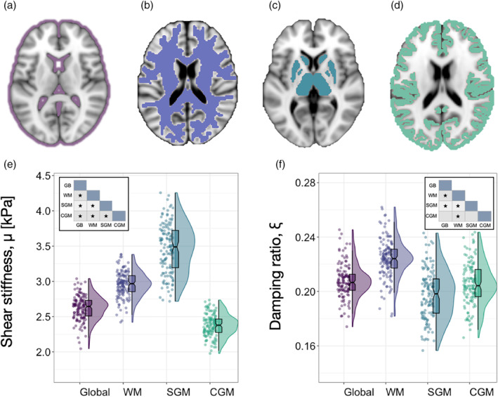
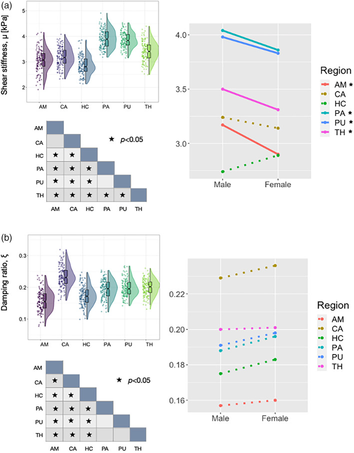
Standard anatomical atlases are common in neuroimaging because they facilitate data analyses and comparisons across subjects and studies. The purpose of this study was to develop a standardized human brain atlas based on the physical mechanical properties (i.e., tissue viscoelasticity) of brain tissue using magnetic resonance elastography (MRE). MRE is a phase contrast-based MRI method that quantifies tissue viscoelasticity noninvasively and in vivo thus providing a macroscopic representation of the microstructural constituents of soft biological tissue. The development of standardized brain MRE atlases are therefore beneficial for comparing neural tissue integrity across populations. Data from a large number of healthy, young adults from multiple studies collected using common MRE acquisition and analysis protocols were assembled (N = 134; 78F/ 56 M; 18-35 years). Nonlinear image registration methods were applied to normalize viscoelastic property maps (shear stiffness, μ, and damping ratio, ξ) to the MNI152 standard structural template within the spatial coordinates of the ICBM-152. We find that average MRE brain templates contain emerging and symmetrized anatomical detail. Leveraging the substantial amount of data assembled, we illustrate that subcortical gray matter structures, white matter tracts, and regions of the cerebral cortex exhibit differing mechanical characteristics. Moreover, we report sex differences in viscoelasticity for specific neuroanatomical structures, which has implications for understanding patterns of individual differences in health and disease. These atlases provide reference values for clinical investigations as well as novel biophysical signatures of neuroanatomy. The templates are made openly available (github.com/mechneurolab/mre134) to foster collaboration across research institutions and to support robust cross-center comparisons.
标准解剖图谱在神经影像学中很常见,因为它们有助于对受试者和研究进行数据分析和比较。本研究旨在使用磁共振弹性成像 (MRE) 基于脑组织的物理力学特性(即组织粘弹性)开发标准化的人脑图谱。MRE 是一种基于相位对比的 MRI 方法,可无创、在体地量化组织粘弹性,从而提供软生物组织微观结构成分的宏观表示。因此,开发标准化的大脑 MRE 图谱有利于比较不同人群的神经组织完整性。使用常见的 MRE 采集和分析方案从多个研究中收集了大量健康年轻成年人的数据(N = 134;78F/56M;18-35 岁)。应用非线性图像配准方法将粘弹性属性图(剪切刚度μ和阻尼比ξ)在空间坐标内归一化为 MNI152 标准结构模板到 ICBM-152。我们发现,平均 MRE 大脑模板包含新出现和对称的解剖细节。利用组装的大量数据,我们表明皮质下灰质结构、白质束和大脑皮层区域表现出不同的力学特性。此外,我们报告了特定神经解剖结构的粘弹性的性别差异,这对理解健康和疾病个体差异的模式具有重要意义。这些图谱为临床研究提供了参考值,以及神经解剖学的新生物物理特征。模板在 github.com/mechneurolab/mre134 上公开提供,以促进研究机构之间的合作,并支持跨中心的稳健比较。