Carle Neuroscience Institute, Carle Foundation Hospital, Urbana, IL, United States; Beckman Institute for Advanced Science and Technology, University of Illinois at Urbana-Champaign, Urbana, IL, United States; Department of Molecular and Integrative Physiology, University of Illinois at Urbana-Champaign, Urbana, IL, United States; Neuroscience Program, University of Illinois at Urbana-Champaign, Urbana, IL, United States.
Beckman Institute for Advanced Science and Technology, University of Illinois at Urbana-Champaign, Urbana, IL, United States; Interdisciplinary Health Sciences Institute, University of Illinois at Urbana-Champaign, Urbana, IL, United States.
Neuroimage Clin. 2020;27:102313. doi: 10.1016/j.nicl.2020.102313. Epub 2020 Jun 16.
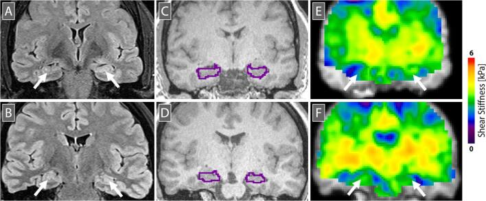
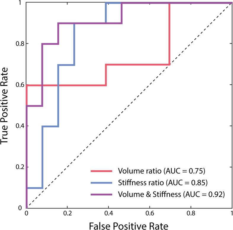
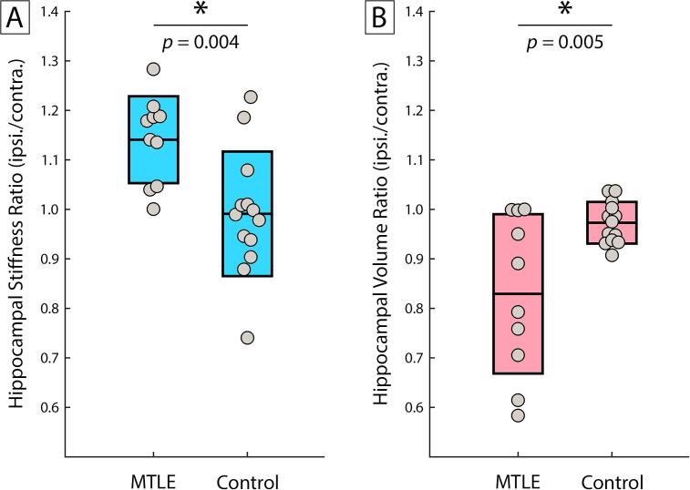
Mesial temporal lobe epilepsy (MTLE) is the most common form of refractory epilepsy. Common imaging biomarkers are often not sensitive enough to identify MTLE sufficiently early to facilitate the greatest benefit from surgical or pharmacological intervention. The objective of this work is to establish hippocampal stiffness measured with magnetic resonance elastography (MRE) as a biomarker for MTLE; we hypothesized that the epileptogenic hippocampus in MTLE is stiffer than the non-epileptogenic hippocampus. MRE was used to measure hippocampal stiffness in a group of patients with unilateral MTLE (n = 12) and a group of healthy comparison participants (n = 13). We calculated the ratio of hippocampal stiffness ipsilateral to epileptogenesis to the contralateral side for both groups. We found a higher hippocampal stiffness ratio in patients with MTLE compared with healthy participants (1.14 v. 0.99; p = 0.004), and that stiffness ratio differentiated MTLE from control groups effectively (AUC = 0.85). Hippocampal stiffness ratio, when added to volume ratio, an established MTLE biomarker, significantly improved the ability to differentiate the two groups (p = 0.038). Stiffness measured with MRE is sensitive to hippocampal pathology in MTLE and the addition of MRE to neuroimaging assessments may improve detection and characterization of the disease.
内侧颞叶癫痫(MTLE)是最常见的难治性癫痫形式。常见的影像学生物标志物通常不够敏感,无法足够早地识别 MTLE,从而无法从手术或药物干预中获得最大益处。这项工作的目的是确定磁共振弹性成像(MRE)测量的海马体硬度是否可作为 MTLE 的生物标志物;我们假设 MTLE 中的致痫海马体比非致痫海马体更硬。MRE 用于测量一组单侧 MTLE 患者(n=12)和一组健康对照组参与者(n=13)的海马体硬度。我们计算了两组的同侧致痫性到对侧的海马体硬度比。我们发现 MTLE 患者的海马体硬度比健康参与者更高(1.14 比 0.99;p=0.004),并且硬度比能够有效地将 MTLE 与对照组区分开(AUC=0.85)。海马体硬度比与体积比(一种已建立的 MTLE 生物标志物)相加后,显著提高了区分两组的能力(p=0.038)。MRE 测量的硬度对 MTLE 中的海马体病理学敏感,将 MRE 添加到神经影像学评估中可能会提高对疾病的检测和特征描述。