Institute of Cell Biology, Heussler Research Group, University of Bern, Bern, Switzerland.
Center for Infectious Diseases, Integrative Parasitology, Heidelberg University Medical School, Heidelberg, Germany.
PLoS One. 2020 Sep 16;15(9):e0238134. doi: 10.1371/journal.pone.0238134. eCollection 2020.
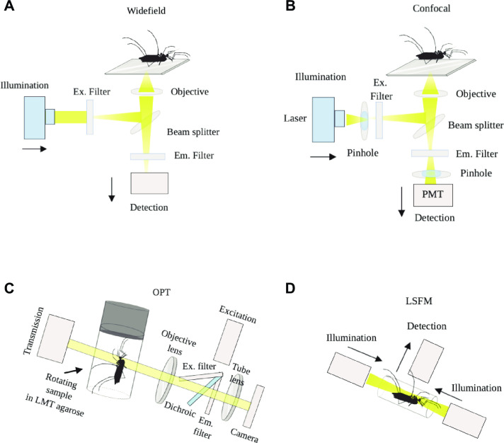
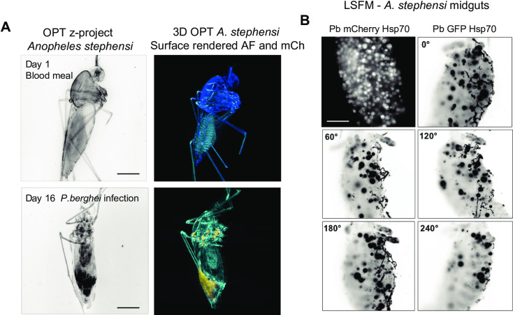
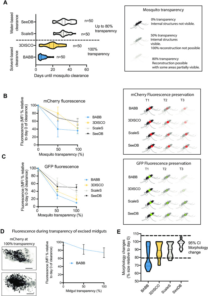
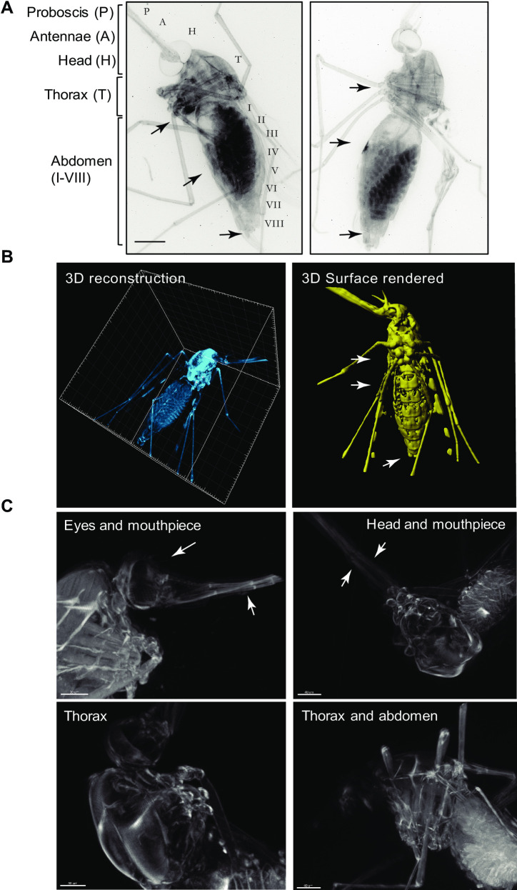
Malaria is a life-threatening disease, caused by Apicomplexan parasites of the Plasmodium genus. The Anopheles mosquito is necessary for the sexual replication of these parasites and for their transmission to vertebrate hosts, including humans. Imaging of the parasite within the insect vector has been attempted using multiple microscopy methods, most of which are hampered by the presence of the light scattering opaque cuticle of the mosquito. So far, most imaging of the Plasmodium mosquito stages depended on either sectioning or surgical dissection of important anatomical sites, such as the midgut and the salivary glands. Optical projection tomography (OPT) and light sheet fluorescence microscopy (LSFM) enable imaging fields of view in the centimeter scale whilst providing micrometer resolution. In this paper, we compare different optical clearing protocols and present reconstructions of the whole body of Plasmodium-infected, optically cleared Anopheles stephensi mosquitoes and their midguts. The 3D-reconstructions from OPT imaging show detailed features of the mosquito anatomy and enable overall localization of parasites in midguts. Additionally, LSFM imaging of mosquito midguts shows detailed distribution of oocysts in extracted midguts. This work was submitted as a pre-print to bioRxiv, available at https://www.biorxiv.org/content/10.1101/682054v2.
疟疾是一种危及生命的疾病,由疟原虫属的顶复门寄生虫引起。疟蚊对这些寄生虫的有性繁殖和向脊椎动物宿主(包括人类)的传播是必要的。已经尝试使用多种显微镜方法对昆虫媒介中的寄生虫进行成像,其中大多数方法都受到蚊子光散射不透明外骨骼的阻碍。到目前为止,对疟蚊阶段的大多数成像都依赖于对重要解剖部位(如中肠和唾液腺)进行切片或手术解剖。光学投影断层扫描(OPT)和光片荧光显微镜(LSFM)能够在厘米范围内成像视场,同时提供微米级分辨率。在本文中,我们比较了不同的光学透明化方案,并展示了感染疟原虫的光学透明化的按蚊斯蒂芬斯蚊子及其中肠的整个身体的重建。来自 OPT 成像的 3D 重建显示了蚊子解剖结构的详细特征,并能够在中肠中对寄生虫进行整体定位。此外,LSFM 对蚊子中肠的成像显示了提取的中肠中卵囊的详细分布。这项工作作为预印本提交给了 bioRxiv,可在 https://www.biorxiv.org/content/10.1101/682054v2 上查看。