Institute of Cell Biology, Heussler Group, University of Bern, Bern, Switzerland.
Wellcome Centre for Integrative Parasitology, University of Glasgow, Glasgow, UK.
Cell Microbiol. 2019 May;21(5):e13023. doi: 10.1111/cmi.13023. Epub 2019 Apr 3.
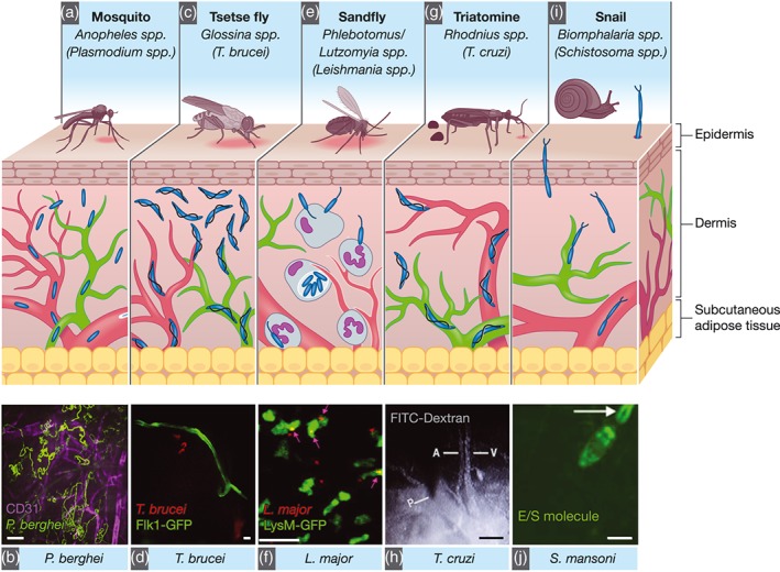
Intravital microscopy allows the visualisation of how pathogens interact with host cells and tissues in living animals in real time. This method has enabled key advances in our understanding of host-parasite interactions under physiological conditions. A combination of genetics, microscopy techniques, and image analysis have recently facilitated the understanding of biological phenomena in living animals at cellular and subcellular resolution. In this review, we summarise findings achieved by intravital microscopy of the skin and adipose tissues upon infection with various parasites, and we present a view into possible future applications of this method.
活体显微镜使我们能够实时观察病原体在活体动物中与宿主细胞和组织的相互作用。这种方法使我们能够在生理条件下深入了解宿主-寄生虫相互作用。遗传学、显微镜技术和图像分析的结合最近促进了我们对活体动物中细胞和亚细胞水平上的生物学现象的理解。在这篇综述中,我们总结了活体显微镜在各种寄生虫感染皮肤和脂肪组织时的研究结果,并展望了该方法的未来应用。