Department of Onco-Hematology, Cell and Gene Therapy, Bambino Gesù Children's Hospital-IRCCS, 00146 Rome, Italy.
Confocal Microscopy Core Facility, Research Center, Bambino Gesù Children's Hospital-IRCCS, 00146 Rome, Italy.
Int J Mol Sci. 2020 Sep 15;21(18):6763. doi: 10.3390/ijms21186763.
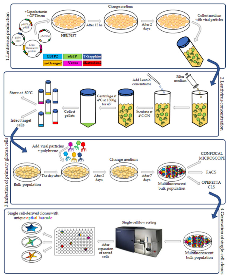
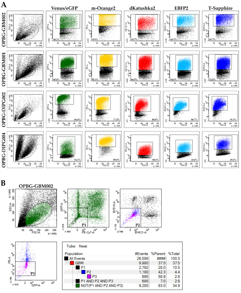
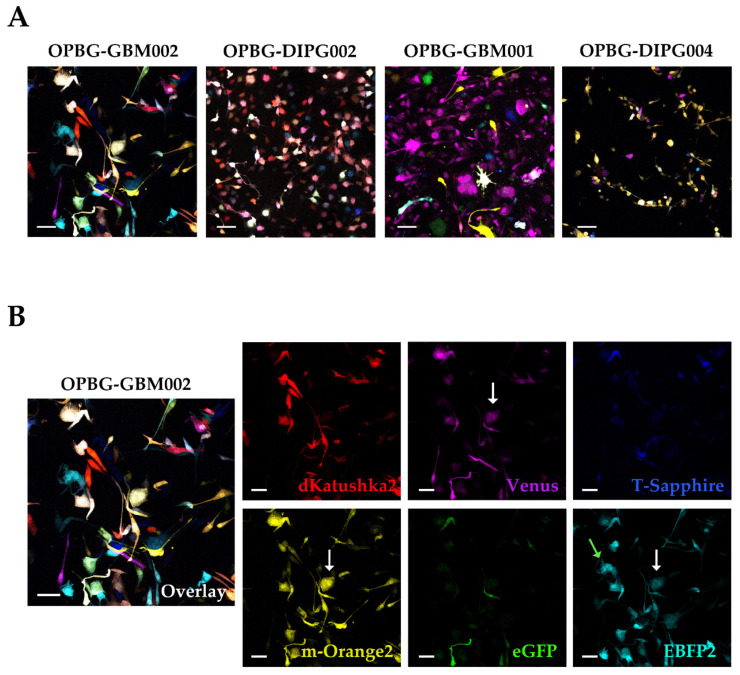
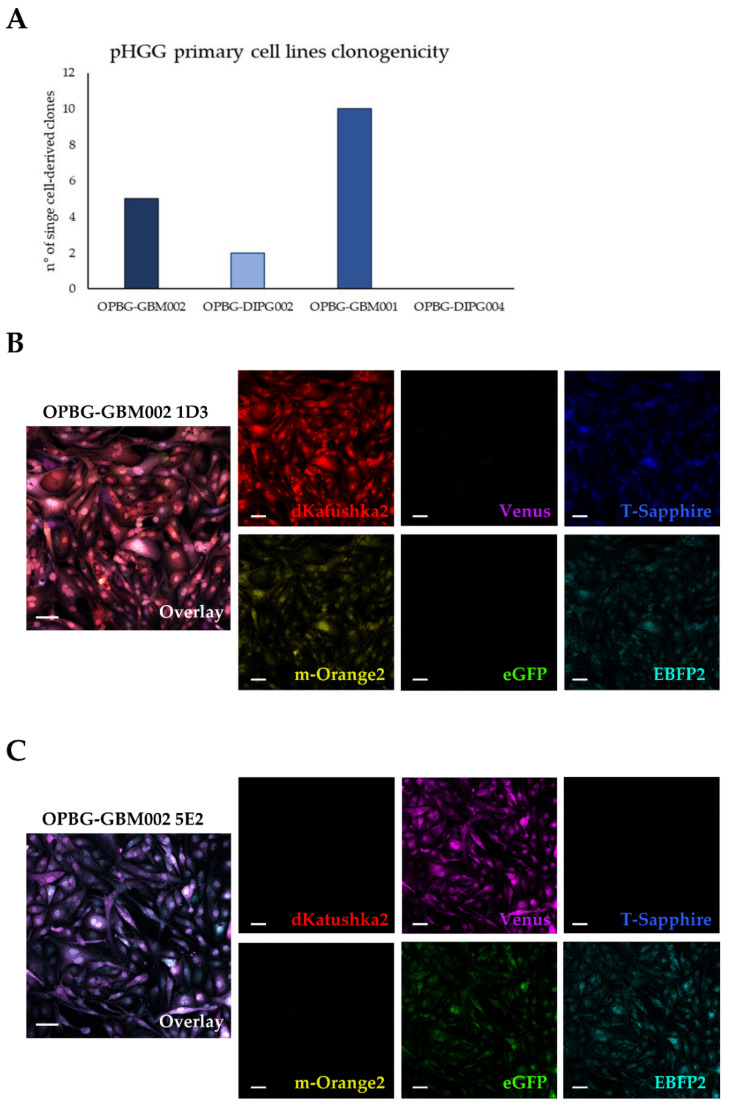
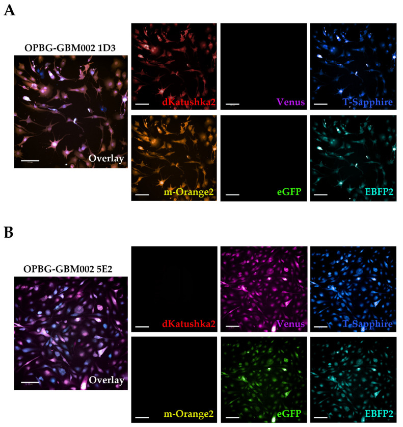
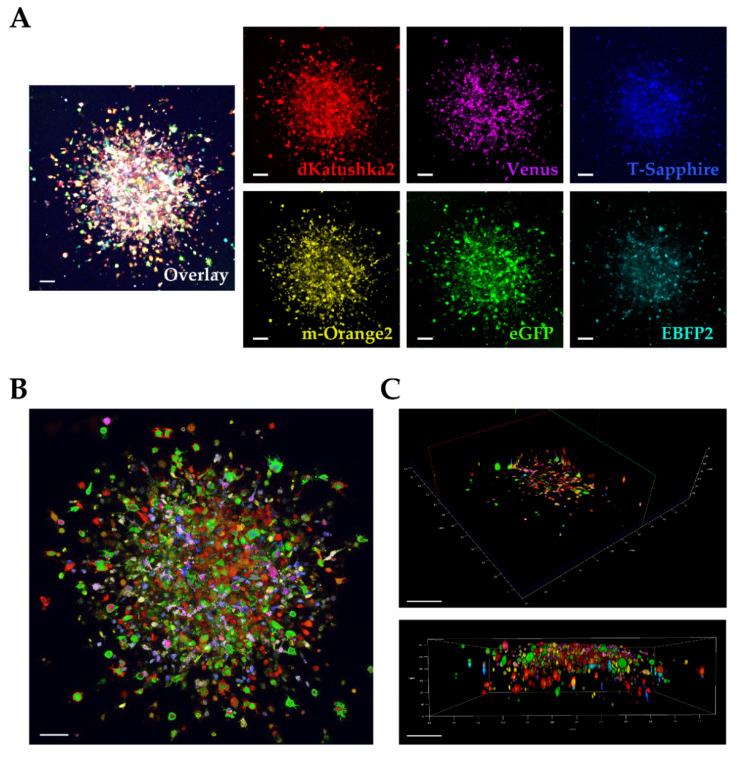
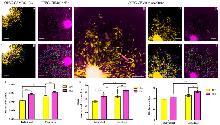
The intratumor heterogeneity represents one of the most difficult challenges for the development of effective therapies to treat pediatric glioblastoma (pGBM) and diffuse intrinsic pontine glioma (DIPG). These brain tumors are composed of heterogeneous cell subpopulations that coexist and cooperate to build a functional network responsible for their aggressive phenotype. Understanding the cellular and molecular mechanisms sustaining such network will be crucial for the identification of new therapeutic strategies. To study more in-depth these mechanisms, we sought to apply the Multifluorescent Marking Technology. We generated multifluorescent pGBM and DIPG bulk cell lines randomly expressing six different fluorescent proteins and from which we derived stable optical barcoded single cell-derived clones. In this study, we focused on the application of the Multifluorescent Marking Technology in 2D and 3D in vitro/ex vivo culture systems. We discuss how we integrated different multimodal fluorescence analysis platforms, identifying their strengths and limitations, to establish the tools that will enable further studies on the intratumor heterogeneity and interclonal interactions in pGBM and DIPG.
肿瘤内异质性是开发有效治疗儿科脑胶质瘤(pGBM)和弥漫性内在脑桥胶质瘤(DIPG)方法的最困难挑战之一。这些脑肿瘤由异质性细胞亚群组成,这些细胞亚群共存并合作构建一个负责其侵袭表型的功能网络。了解维持这种网络的细胞和分子机制对于确定新的治疗策略至关重要。为了更深入地研究这些机制,我们试图应用多荧光标记技术。我们生成了随机表达六种不同荧光蛋白的多荧光 pGBM 和 DIPG 细胞系,并从中衍生出稳定的光学条形码单细胞衍生克隆。在这项研究中,我们专注于多荧光标记技术在 2D 和 3D 体外/体外培养系统中的应用。我们讨论了如何整合不同的多模态荧光分析平台,确定它们的优势和局限性,以建立能够进一步研究 pGBM 和 DIPG 肿瘤内异质性和克隆间相互作用的工具。