Huang Yiyao, Cheng Lesley, Turchinovich Andrey, Mahairaki Vasiliki, Troncoso Juan C, Pletniková Olga, Haughey Norman J, Vella Laura J, Hill Andrew F, Zheng Lei, Witwer Kenneth W
Department of Molecular and Comparative Pathobiology, Johns Hopkins University School of Medicine, Baltimore, MD, USA.
Department of Laboratory Medicine, Nanfang Hospital, Southern Medical University, Guangzhou, Guangdong, China.
J Extracell Vesicles. 2020 Jun 30;9(1):1785746. doi: 10.1080/20013078.2020.1785746.
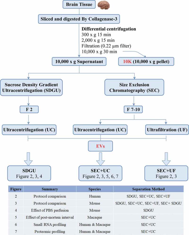
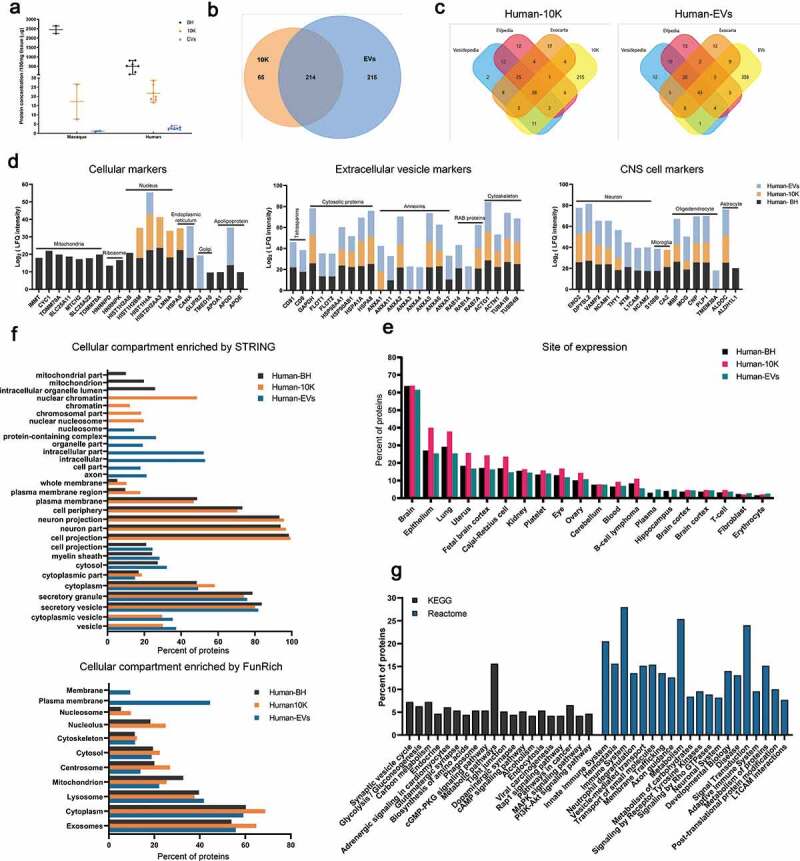
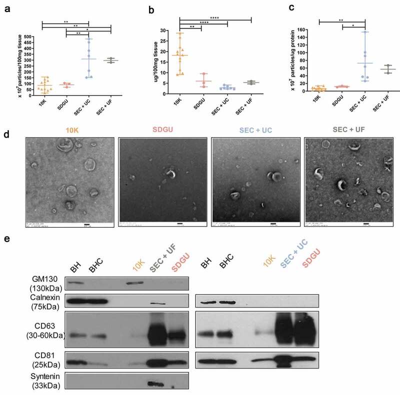
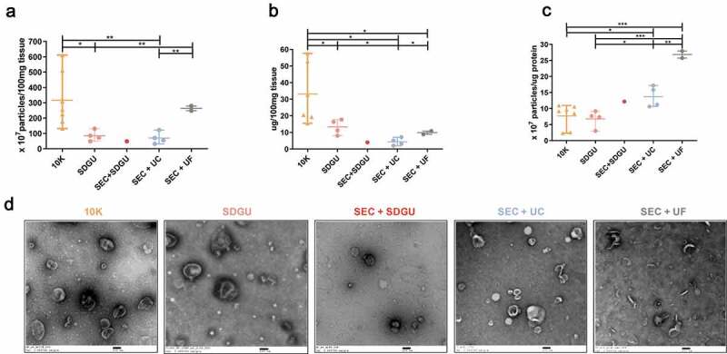
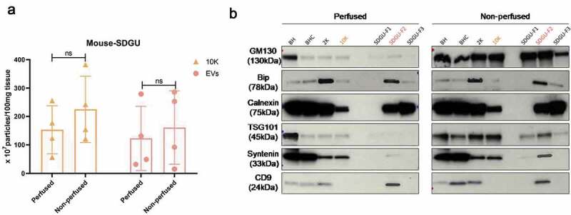
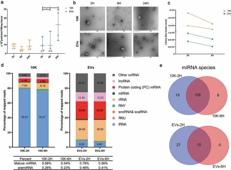
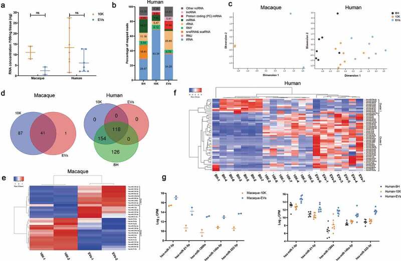
Extracellular vesicles (EVs) are involved in a wide range of physiological and pathological processes by shuttling material out of and between cells. Tissue EVs may thus lend insights into disease mechanisms and also betray disease when released into easily accessed biological fluids. Since brain-derived EVs (bdEVs) and their cargo may serve as biomarkers of neurodegenerative diseases, we evaluated modifications to a published, rigorous protocol for separation of EVs from brain tissue and studied effects of processing variables on quantitative and qualitative outcomes. To this end, size exclusion chromatography (SEC) and sucrose density gradient ultracentrifugation were compared as final separation steps in protocols involving stepped ultracentrifugation. bdEVs were separated from brain tissues of human, macaque, and mouse. Effects of tissue perfusion and a model of post-mortem interval (PMI) before final bdEV separation were probed. MISEV2018-compliant EV characterization was performed, and both small RNA and protein profiling were done. We conclude that the modified, SEC-employing protocol achieves EV separation efficiency roughly similar to a protocol using gradient density ultracentrifugation, while decreasing operator time and, potentially, variability. The protocol appears to yield bdEVs of higher purity for human tissues compared with those of macaque and, especially, mouse, suggesting opportunities for optimization. Where possible, perfusion should be performed in animal models. The interval between death/tissue storage/processing and final bdEV separation can also affect bdEV populations and composition and should thus be recorded for rigorous reporting. Finally, different populations of EVs obtained through the modified method reported herein display characteristic RNA and protein content that hint at biomarker potential. To conclude, this study finds that the automatable and increasingly employed technique of SEC can be applied to tissue EV separation, and also reveals more about the importance of species-specific and technical considerations when working with tissue EVs. These results are expected to enhance the use of bdEVs in revealing and understanding brain disease.
细胞外囊泡(EVs)通过在细胞间穿梭物质参与广泛的生理和病理过程。因此,组织来源的EVs可能有助于深入了解疾病机制,并且当它们释放到易于获取的生物体液中时也能揭示疾病。由于脑源性EVs(bdEVs)及其所载物质可能作为神经退行性疾病的生物标志物,我们评估了对已发表的、从脑组织中分离EVs的严格方案的改进,并研究了处理变量对定量和定性结果的影响。为此,在涉及分步超速离心的方案中,比较了尺寸排阻色谱(SEC)和蔗糖密度梯度超速离心作为最终分离步骤。从人、猕猴和小鼠的脑组织中分离bdEVs。探讨了组织灌注和最终bdEV分离前的死后间隔(PMI)模型的影响。进行了符合MISEV2018标准的EV表征,并对小RNA和蛋白质进行了分析。我们得出结论,改进后的采用SEC的方案实现的EV分离效率与使用梯度密度超速离心的方案大致相似,同时减少了操作人员的时间,并可能降低变异性。与猕猴尤其是小鼠的组织相比,该方案似乎能为人脑组织产生更高纯度的bdEVs,这表明有优化的机会。在可能的情况下,应在动物模型中进行灌注。死亡/组织储存/处理与最终bdEV分离之间的间隔也会影响bdEV的群体和组成,因此应记录下来以便进行严格的报告。最后,通过本文报道的改进方法获得的不同群体的EVs显示出特征性的RNA和蛋白质含量,暗示了其作为生物标志物的潜力。总之,本研究发现可自动化且越来越常用的SEC技术可应用于组织EV分离,并且还揭示了在处理组织EVs时物种特异性和技术考虑因素的重要性。这些结果有望加强bdEVs在揭示和理解脑部疾病中的应用。